. 2018 Sep 1;28(9):3255-3266.
doi: 10.1093/cercor/bhx196.

Neuron-Targeted Caveolin-1 Promotes Ultrastructural and Functional Hippocampal Synaptic Plasticity

Affiliations

Neuron-Targeted Caveolin-1 Promotes Ultrastructural and Functional Hippocampal Synaptic Plasticity

Junji Egawa et al. Cereb Cortex. .

Abstract

A delicate interneuronal communication between pre- and postsynaptic membranes is critical for synaptic plasticity and the formation of memory. Evidence shows that membrane/lipid rafts (MLRs), plasma membrane microdomains enriched in cholesterol and sphingolipids, organize presynaptic proteins and postsynaptic receptors necessary for synaptic formation and signaling. MLRs establish a cell polarity that facilitates transduction of extracellular cues to the intracellular environment. Here we show that neuron-targeted overexpression of an MLR protein, caveolin-1 (SynCav1), in the adult mouse hippocampus increased the number of presynaptic vesicles per bouton, total excitatory type I glutamatergic synapses, number of same-dendrite multiple-synapse boutons, increased myelination, increased long-term potentiation, and increased MLR-localized N-methyl-d-aspartate receptor subunits (GluN1, GluN2A, and GluN2B). Immunogold electron microscopy revealed that Cav-1 localizes to both the pre- and postsynaptic membrane regions as well as in the synaptic cleft. These findings, which are consistent with a significant increase in ultrastructural and functional synaptic plasticity, provide a fundamental framework that underlies previously demonstrated improvements in learning and memory in adult and aged mice by SynCav1. Such observations suggest that Cav-1 and MLRs alter basic aspects of synapse biology that could serve as potential therapeutic targets to promote neuroplasticity and combat neurodegeneration in a number of neurological disorders.


Figures

Figure 1.
Figure 1.
SynCav1 increases total number of synapses and PSVs in the hippocampus. AAV9-SynRFP or AAV9-SynCav1 was delivered to the hippocampus using stereotaxis, and 2 months later brains were prepared for routine EM. Hippocampal CA1 distal apical dendrites in the “stratum radiatum” were examined for ultrastructural changes using EM. (Ai,ii) SynRFP-injected mice; (Bi,ii) SynCav1-injected mice. a, axons; d, dendritic spines; asterisks indicate multiple-synapse boutons as previously described (Geinisman 1993; Fiala et al. 2002). Quantification of total synapses (C) and PSVs/axonal bouton (D). “Left panels” (9300× Mag); “right panels” (13 000× Mag of inset in “left panels”). (*P < 0.05, n = 3/group). Scale bar = 500 nm. (E) Hippocampal schematic illustrating the CA1 dendritic region (orange rectangle) used for EM imaging.
Figure 2.
Figure 2.
SynCav1 increases total number of dsMBSs in the hippocampus. AAV9-SynRFP or AAV9-SynCav1 was delivered directly to the hippocampus using stereotaxis, and 2 months later brains were prepared for routine EM. CA1 apical region in the hippocampus was examined for ultrastructural changes. (A) SynRFP-injected mice; examples of sdMSBs (Geinisman 1993; Fiala et al. 2002) are shown in insets i and ii. (B) SynCav1-injected mice; examples of sdMSBs are shown in insets i–iv; arrow head in Bii indicates polyribosome. “Left panels” (4800× Mag); “insets” (2.5× of 4800× image). (C) Quantification of A and B (*P < 0.05, n = 3/group). Scale bar = 1 μm; inset scale bar = 0.25 μm.
Figure 3.
Figure 3.
SynCav1 increases myelin structure in the hippocampus. AAV9-SynRFP or AAV9-SynCav1 was delivered directly to the hippocampus using stereotaxis, and 2 months later brains were prepared for routine EM. G-ratio analysis (axon lumen diameter/fiber diameter) was used to measure changes in myelination in Schaffer axons innervating the CA1 apical dendritic arbor in the hippocampus. (Ai) 4800× Mag of SynRFP-injected mice; (Aii) 3.5× magnification of the inset shown in (Ai). (Bi) 4800× Mag of SynCav1-injected mice; (Bii) 3.5× magnification of the inset shown in (Bi). Quantification of G-ratio (C), axonal lumen diameter (D), and myelin sheath diameter (E) was conducted using Adobe Photoshop (*P < 0.05, n = 3/group). Scale bar = 1 μm.
Figure 4.
Figure 4.
Immunogold EM showing Cav-1 subsynaptic localization at excitatory asymmetric synapses on distal apical CA1 dendrites. AAV9-SynCav1 (or AAV9-SynRFP) was delivered to the hippocampus using stereotaxis, and 2 months later brains were prepared for immunogold EM. Hippocampal CA1 distal apical dendrites in the “stratum radiatum” were examined for subsynaptic localization of Cav-1 (12 nm gold) along with the PS markers SYNPHYS (18 nm gold) and Synap (18 nm) or the postsynaptic receptor TrkB (18 nm gold). Immunogold EM images are show in (Ai–iii) SynRFP and in (Bi–iii) SynCav1. Quantitation of total Cav-1 immunogold particles in PS (C) and postsynaptic (D) regions normalized per synapse. Distribution of Cav-1 immunogold particles relative to PS (E) and postsynaptic (F) membranes normalized per synapse. Morphometric analysis for PSD length (G), width (H), area (I), and perimeter (J). Data (mean ± SEM) were normalized to synapse as indicated in Y-axis (****P < 0.0001; ***P = 0.0001; **P = 0.002; *P = 0.02). Scale bar = 200 nm.
Figure 5.
Figure 5.
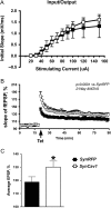
Effects of SynCav1 and SynRFP on electrophysiological properties of the Schaffer collateral/CA1 synaptic pathway. (A) Input–output dependencies were similar in hippocampal slices prepared from AAV9-SynCav1 and AAV9-SynRFP-treated mice. (B) LTP recording of field responses in the CA1 was measured over time following tetanization of the CA3 Schaffer collaterals. Tetanizations of afferent fibers (Tet, arrow) resulted in robust and stable increases of the evoked responses in both SynCav1 and SynRFP mice. Note that the responses were greater in the SynCav1-injected mice. (C) Mean values of LTP shown in (B) (*P < 0.05, n = 8–10/group). LTP values shown in C represents averages taken 50–70 min after tetanus.
Figure 6.
Figure 6.
SynCav1 significantly enhances MLR-localization of NMDAR subunits in adult hippocampus. (A) Hippocampal (Hpc) homogenates from AAV9-SynCav1 and AAV9-SynRFP-treated mice were immunobloted for NMDAR subunits GluN1A, GluN2A, GluN2B, and Cav-1. Quantitation (normalized to GAPDH) is shown in (B). (C) Hpc homogenates (equal protein loading of 0.5 mg/mL) were subjected to sucrose density fraction (BFs) followed by immunoblot analysis. Quantitation is shown in (D). Red box demarcates MLR fractions 4 and 5 (BF). Data represent arbitrary units (A.U.) mean ± SEM (*P < 0.05, n = 4/group).

References

    1. Abraham WC. 2003. How long will long-term potentiation last? Philos Trans R Soc Lond B Biol Sci. 358:735–744. - PMC - PubMed
    1. Andersen P. 1960. Interhippocampal impulses. II. Apical dendritic activation of CAI neurons. Acta Physiol Scand. 48:178–208. - PubMed
    1. Andersen P, Blackstad TW, Lomo T. 1966. Location and identification of excitatory synapses on hippocampal pyramidal cells. Exp Brain Res. 1:236–248. - PubMed
    1. Andersen P, Morris R, Amaral D, Bliss T, O’Keefe J. 2007. The Hippocampus Book. Oxford Neuroscience Series. New York:Oxford University Press.
    1. Bengtsson SL, Nagy Z, Skare S, Forsman L, Forssberg H, Ullen F. 2005. Extensive piano practicing has regionally specific effects on white matter development. Nat Neurosci. 8:1148–1150. - PubMed

Publication types