. 2017 Nov 2;45(19):10948-10968.
doi: 10.1093/nar/gkx805.

Embraced by eIF3: structural and functional insights into the roles of eIF3 across the translation cycle

Affiliations

Embraced by eIF3: structural and functional insights into the roles of eIF3 across the translation cycle

Leoš Shivaya Valášek et al. Nucleic Acids Res. .

Abstract

Protein synthesis is mediated via numerous molecules including the ribosome, mRNA, tRNAs, as well as translation initiation, elongation and release factors. Some of these factors play several roles throughout the entire process to ensure proper assembly of the preinitiation complex on the right mRNA, accurate selection of the initiation codon, errorless production of the encoded polypeptide and its proper termination. Perhaps, the most intriguing of these multitasking factors is the eukaryotic initiation factor eIF3. Recent evidence strongly suggests that this factor, which coordinates the progress of most of the initiation steps, does not come off the initiation complex upon subunit joining, but instead it remains bound to 80S ribosomes and gradually falls off during the first few elongation cycles to: (1) promote resumption of scanning on the same mRNA molecule for reinitiation downstream-in case of translation of upstream ORFs short enough to preserve eIF3 bound; or (2) come back during termination on long ORFs to fine tune its fidelity or, if signaled, promote programmed stop codon readthrough. Here, we unite recent structural views of the eIF3-40S complex and discus all known eIF3 roles to provide a broad picture of the eIF3's impact on translational control in eukaryotic cells.


Figures

Figure 1.
Figure 1.
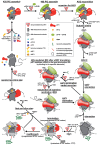
Schematics of the entire translational cycle with ‘detours’ for: (1) reinitiation, (2) programmed stop codon readthrough and (3) the Nonsense-mediated decay pathway, highlighting the role of eIF3 at the individual steps. For details, see the main text.
Figure 2.
Figure 2.
Schematics of the S. cerevisiae eIF3 complex. A 3D model of yeast eIF3 and its associated eIFs in the Multifactor complex (MFC) composed together to the best possible fit from all available structures of individual domains; namely the X-ray structure of the yeast i/TIF34 (full length)—g/TIF35-NTD (S.c. residues 1–135)—b/PRT1-extended-α-helix (S.c. residues 655–698) complex, the X-ray structure of the β-propeller (formed by nine WD40 repeats) of the middle domain of b/PRT1 (residues 132–626), the X-ray structure of the mutually interacting PCI domains of a/TIF32 (residues 1–496) and c/NIP1 (residues 251–812) (all taken from (64)); the NMR structure of the interaction between the RRM of human eIF3b (H.s. residues 170–274) and the N-terminal peptide of human eIF3j (H.s. residues 35–69) (37), and the NMR structure of the C-terminal RRM of human eIF3g (H.s. residues 231–320) (38). Arrows indicate all known interactions of eIF3 domains with other eIFs, ribosomal proteins and mRNA (see text for further details). NTD, N-terminal domain; CTD, C-terminal domain; HLD, HCR1-like domain; RRM, RNA recognition motif; PCI, PCI domain; WD40, WD40 domain; TC, ternary complex (composed of eIF2•GTP•Met-tRNAiMet).
Figure 3.
Figure 3.
Schematics of the human eIF3 complex. (A) A schematic model of human eIF3 was adapted from (25). The eIF3 subunits forming the PCI/MPN octamer with the anthropomorphic shape are indicated by the grey background. The rectangle marks the seven α-helices involved in formation of the 7-helix bundle shown in panel C. The Yeast-Like Core (YLC) comprising the eIF3 subunits a, b, g and i (defined previously by (41)) is depicted and so is the eIF3-associated factor eIF3j with arrows indicating its contacts with other eIF3 subunits. The upper right-hand side arrow indicates the interaction between eIF3e and eIF3d that attaches eIF3d to the rest of eIF3 (48,59,159). Arrows indicate all known interactions of eIF3 domains with other eIFs, ribosomal proteins and mRNA (see text for further details). (B) Polyalanine-level model of the eIF3 octamer core with the close-up view of the 7-helix bundle formed by subunits h, c, e, f, l and k (adapted from (59)).
Figure 4.
Figure 4.
Schematic representation of the arrangement of eIF3 subunits in the two available conformations that are deduced from the available Cryo-EM analysis (adapted from (59)). (A) eIF3 binds to the solvent-exposed side with the octamer occupying the platform of the small ribosomal subunit connected with the eIF3b–g–i module (YLC)—sitting near the mRNA entry channel—via the extended C-terminal linker domain of eIF3a (a dashed red line indicates a predicted location of the eIF3a-CTD; placement of the eIF3g-RRM is also only our best guess) (,–65). Figure includes only those domains of eIF3 subunits for which the structures are known. The 40S subunit is depicted in grey surface; all other subunits are labelled and colored variably. The eIF3 helical bundles fortifying the intersubunit interactions are represented as cylinders. The predicted path of mRNA is shown in dark red; Ex and En—mRNA exit and entry channels, respectively. For details please see the main text. (B) In this conformation, the entire eIF3a-CTD–b–g–i module relocates from the solvent-exposed side to the intersubunit side, so that the eIF3b-RRM interacts with 18S rRNA and eIF1 and the eIF3b-propeller interacts with eIF2γ (66,70); a density presumably corresponding to the eIF3c-NTD residues 115–220 was in this structure identified not too far away from eIF1, where it could co-ordinate AUG recognition with other eIFs. Purely hypothetically, this could be the conformation that eIF3 adopts upon AUG recognition. Placement of the eIF3g–i unit held by the eIF3b-extended α-helix is only our best guess; for details please see the main text. (C) Atomic model of rabbit eIF3b (RRM, WD40 and the C-terminal extended α-helix domains - orange), yeast eIF3i-WD40 (purple) and a long α-helix (red) corresponding to a fragment of the C-terminal helical region of eIF3a. For details please see the main text.
Figure 5.
Figure 5.
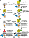
Translation initiation factor eIF3 promotes programmed stop codon readthrough - revised model adapted from (21). (A and B) Canonical termination; stop codon in the termination favorable context appears in the A-site (only UAG and UAA stop codons are indicated for illustration purposes; UGA works by the same mechanism; +4 base is indicated by a question mark), eRF1 in complex with eRF3.GTP binds to it and samples the codon in a two-step process (100). In the first step, the first and second nucleotides of the stop codon are recognized by specific residues of the eRF1-NTD. This is followed by the eRF1-NTD conformational re-arrangement during the second step, which probably includes flipping the A1493 base (according to the E. coli nomenclature) accompanied by formation of the U-turn-like conformation (103,104). This step permits decoding of the third nucleotide. As a result, eRF1 stably accommodates in the A-site triggering GTP hydrolysis on eRF3, followed by polypeptide release and ribosomal recycling. eIF3 has minimal, if any role here (see text for further details). (C and D) Programmed stop codon readthrough; stop codon occurs in the unfavorable termination context bearing specific consensus sequences like CAR-NBA in its 3′ UTR—in this particular case proposed to base-pair with 18S rRNA (160). The eIF3 presence in the pre-termination complex—perhaps in co-operation with these sequences—prevents the A1493 phosphate group to flip and thus specifically interferes with the proper decoding of the third position of programmed stop codons (19,21). This results in ejection of the eRF1–eRF3•GTP complex from the pre-termination complexes allowing incorporation of near-cognate tRNAs with the mismatch at the third position to read through the stop codon and continue with elongation. For details please see the main text.
Figure 6.
Figure 6.
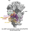
Graphical illustration (adapted from (47)) of the proposed arrangement of the post-termination complex on uORF1 with its RPEs interacting with Box 6 and Box 17 segments of the N-terminal domain of a/TIF32 to promote resumption of scanning for REI on GCN4. The exit channel view of the py48S-closed complex shows only two incomplete eIF3 subunits for simplicity: c/NIP1 in wheat and a/TIF32 in purple with its extreme NTD in light purple and its C-terminal HCR1-like domain (HLD) represented by a dotted line (its structure is unknown and thus its placement in the py48S complex was only predicted). The location of both a/TIF32 boxes is marked in green; the 5′-UTR of uORF1 is shown in orange with its RPEs depicted in yellow-orange.

Similar articles

Cited by

References

    1. Hinnebusch A.G. Structural insights into the mechanism of scanning and start codon recognition in eukaryotic translation initiation. Trends Biochem. Sci. 2017; 42:589–611. - PubMed
    1. Valášek L.S. 'Ribozoomin’—translation initiation from the perspective of the ribosome-bound eukaryotic initiation factors (eIFs). Curr. Protein Pept. Sci. 2012; 13:305–330. - PMC - PubMed
    1. Fringer J.M., Acker M.G., Fekete C.A., Lorsch J.R., Dever T.E.. Coupled release of eukaryotic translation initiation factors 5B and 1A from 80S ribosomes following subunit joining. Mol. Cell Biol. 2007; 27:2384–2397. - PMC - PubMed
    1. Jennings M.D., Kershaw C.J., Adomavicius T., Pavitt G.D.. Fail-safe control of translation initiation by dissociation of eIF2alpha phosphorylated ternary complexes. Elife. 2017; 6:e24542. - PMC - PubMed
    1. Hronova V., Valasek L.S.. An emergency brake for protein synthesis. Elife. 2017; 6:e27085. - PMC - PubMed

MeSH terms

LinkOut - more resources