Review
. 2018 Jan;1412(1):170-177.
doi: 10.1111/nyas.13472. Epub 2017 Oct 5.

Homeostatic synaptic plasticity at the neuromuscular junction in myasthenia gravis

Affiliations
Review

Homeostatic synaptic plasticity at the neuromuscular junction in myasthenia gravis

Xueyong Wang et al. Ann N Y Acad Sci. 2018 Jan.

Abstract

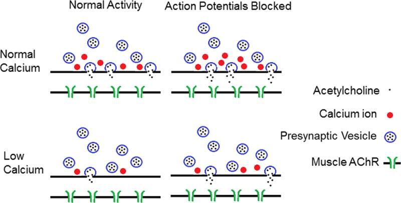
A number of studies in the past 20 years have shown that perturbation of activity of the nervous system leads to compensatory changes in synaptic strength that serve to return network activity to its original level. This response has been termed homeostatic synaptic plasticity. Despite the intense interest in homeostatic synaptic plasticity, little attention has been paid to its role in the prototypic synaptic disease, myasthenia gravis. In this review, we discuss mechanisms that have been shown to mediate homeostatic synaptic plasticity at the mammalian neuromuscular junction. A subset of these mechanisms have been shown to occur in myasthenia gravis. The homeostatic changes occurring in myasthenia gravis appear to involve the presynaptic nerve terminal and may even involve changes in the excitability of motor neurons within the spinal cord. The finding of presynaptic homeostatic synaptic plasticity in myasthenia gravis leads us to propose that changes in the motor unit in myasthenia gravis may be more widespread than previously appreciated.

Keywords: acetylcholine receptor; action potential; activity; endplate; synapse; transmission.


Figures

Figure 1
Figure 1
Shown is a cartoon of homeostatic synaptic plasticity at the mouse NMJ triggered by prolonged blockage of nerve action potentials. In the top row, synaptic function is shown in solution containing normal external Ca. Following blockage of action potentials, there are two changes in synaptic function. The first is an increase in the release of acetylcholine (ACh) during fusion of individual synaptic vesicles (illustrated as increased ACh in the synaptic cleft). This is likely responsible for the increase in the amplitude of spontaneous synaptic currents (miniature endplate currents (MEPCs)). The second change is an increase in entry of Ca into the presynaptic terminal during an action potential. However, when extracellular Ca is normal, this has no significant effect on the number of synaptic vesicles released (quantal content (QC)), as each releasable vesicle is already released with each action potential. The bottom row shows the situation when extracellular Ca is lowered. In this case, Ca entry during the action potential limits QC. When Ca entry is increased following blockage of action potentials, even a small increase in Ca entry triggers a significant increase in QC. In addition, the increase in ACh release from each vesicle also occurs. This combines with the increase in QC to cause a dramatic increase in synaptic strength. This is illustrated in the cartoon as an increase from two to eight ACh molecules in the synaptic cleft. AChR, acetylcholine receptor.
Figure 2
Figure 2
Shown is a cartoon of NMJ homeostatic synaptic plasticity following blockage or destruction of AChRs, as occurs in MG. In the top row, synaptic function is shown in solution containing normal external Ca. Two types of synaptic vesicles are present. The blue vesicles represent normal vesicles that participate in synaptic transmission at baseline. The red vesicles represent a special pool of synaptic vesicles that normally do not play an important role in synaptic transmission. A few vesicles in this pool are released late in the course of the synaptic current (indicated by the long dotted red arrow). Following partial blockage of AChRs, the special pool of synaptic vesicles is released more rapidly (synchronous with the normal pool). In addition, the special pool of vesicles rapidly recycles (indicated by the curved red arrow) to increase QC. Because the special pool of vesicles is so small, it is rapidly depleted during repetitive stimulation. In the bottom row is shown the situation when extracellular Ca is lowered. When extracellular Ca is lowered, the special pool of synaptic vesicles does not participate in synaptic transmission (thus, the pool is not shown in the cartoon). This suggests that this pool requires high levels of Ca entry in order to be mobilized for release. Because this pool does not participate in release, blockage of AChRs does not trigger an increase in QC and thus there is functionally no synaptic plasticity.

Similar articles

Cited by

References

    1. Auerbach A. Activation of endplate nicotinic acetylcholine receptors by agonists. Biochem Pharmacol. 2015;97:601–608. - PMC - PubMed
    1. Del Castillo J, Katz B. Quantal components of the end-plate potential. J Physiol. 1954;124:560–573. - PMC - PubMed
    1. Kavalali ET. The mechanisms and functions of spontaneous neurotransmitter release. Nat Rev Neurosci. 2015;16:5–16. - PubMed
    1. Peled ES, Newman ZL, Isacoff EY. Evoked and spontaneous transmission favored by distinct sets of synapses. Curr Biol. 2014;24:484–493. - PMC - PubMed
    1. Heuser JE, et al. Synaptic vesicle exocytosis captured by quick freezing and correlated with quantal transmitter release. J Cell Biol. 1979;81:275–300. - PMC - PubMed

Publication types