Review
. 2018 Jan 17;9(1):51-72.
doi: 10.1021/acschemneuro.7b00271. Epub 2017 Nov 7.

Glycosylation Changes in Brain Cancer

Affiliations
Review

Glycosylation Changes in Brain Cancer

Lucas Veillon et al. ACS Chem Neurosci. .

Abstract

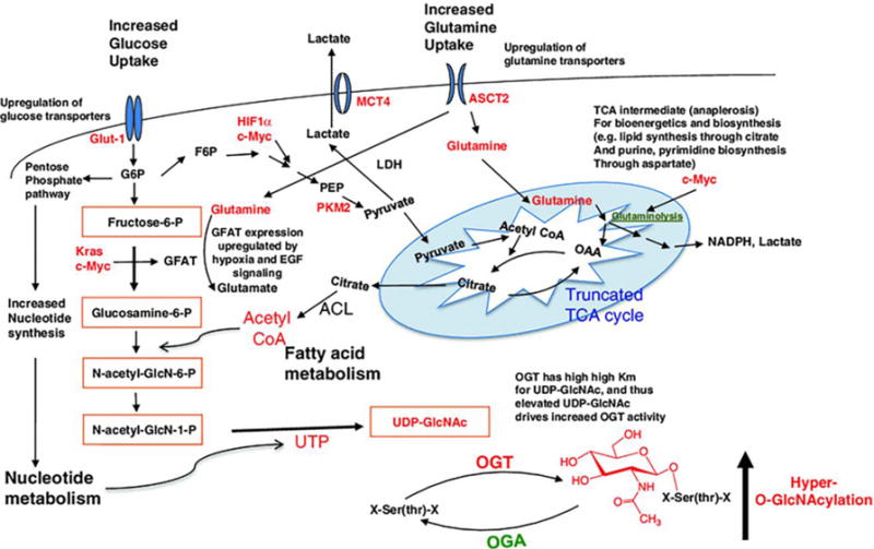
Protein glycosylation is a posttranslational modification that affects more than half of all known proteins. Glycans covalently bound to biomolecules modulate their functions by both direct interactions, such as the recognition of glycan structures by binding partners, and indirect mechanisms that contribute to the control of protein conformation, stability, and turnover. The focus of this Review is the discussion of aberrant glycosylation related to brain cancer. Altered sialylation and fucosylation of N- and O-glycans play a role in the development and progression of brain cancer. Additionally, aberrant O-glycan expression has been implicated in brain cancer. This Review also addresses the clinical potential and applications of aberrant glycosylation for the detection and treatment of brain cancer. The viable roles glycans may play in the development of brain cancer therapeutics are addressed as well as cancer-glycoproteomics and personalized medicine. Glycoprotein alterations are considered as a hallmark of cancer while high expression in body fluids represents an opportunity for cancer assessment.

Keywords: Brain cancer; aberrant glycosylation; bone marrow-derived human mesenchymal stem cells; cancer stem cells; carcinoembryonic antigen; central nervous system; glioblastoma; glioma stem cells; glycosylation; human mucin family; posttranslational modification of proteins; small cell lung carcinomas.


Figures

Figure 1
Figure 1
Glycosylation reactions catalyzed by the glycosyltransferases GnT-III and GnT-V, as well as by Fut8, and their biological functions. Reprinted from, with permission.
Figure 2
Figure 2
In the presence of activating ligands and absence of inhibitory ligands on the target cell, NK cells are activated to release cytotoxic effectors and cytokines. Coating cancer cells with sialylated glycopolymers by membrane insertion can emulate cancer associated glycosylation changes that engage the Siglec family of inhibitory receptors. Localization of Siglecs to the site of activation enhances SHP-1/2 phosphatase recruitment to halt the phosphorylation cascade before cellular activation. Reprinted from, with permission.
Figure 3
Figure 3
Structures of common fucosylated glycans. (A) Synthesis of ABO blood group antigens. The H and Se transferases are a pair of α(1,2)-fucosyltransferases that synthesize the H antigen in a variety of tissues. The ABO locus encodes a glycosyltransferase that further modifies the H antigen. The A allele at the ABO locus encodes an N-acteylgalactosaminyltransferase. The B allele encodes a galactosyltransferase that differs from the A transferase at four amino acid positions. The O allele at the ABO locus encodes a truncated, enzymatically inactive protein. (B) Lewis-related antigens. Circles indicate the immunodominant portion of each antigen. (C) A representative O-linked fucose glycan. Fucose modifies serines or threonine within the broad consensus site shown here, and in Table I. R indicates glycolipid and N- and O-linked glycoprotein precursors. Reprinted from, with permission.
Figure 4
Figure 4
Mucins, chronic inflammation and cancer. In this proposed model of the association of mucins with chronic inflammation and cancer, the production of inflammatory cytokines by immune effector cells activates transcription factors, for example nuclear factor-κB (NF-κB), signal transducer and activator of transcription 1 (STAT1) and STAT3, in epithelial cells. In turn, these transcription factors upregulate mucin expression to enhance the mucous barrier and protect the epithelial layer. Mucin 2 (MUC2) limits the inflammatory response at the apical membrane and inhibits transformation. Upregulation of the MUC1 and MUC4 transmembrane mucins similarly contributes to the protective barrier and loss of polarity in the epithelial stress response. Activation of MUC1 is associated with targeting of the MUC1 C-terminal transmembrane subunit (MUC1-C) to the nucleus, where it promotes a gene programme for proliferation and survival. Targeting of MUC1-C to the mitochondria also blocks cell death to prevent loss of the epithelial barrier. However, with chronic inflammation and prolonged stimulation of this protective response, epithelial cells may become susceptible to the accumulation of genetic mutations that induce transformation in a setting with downregulation of pathways that would otherwise protect against oncogenic events. IL-6, interleukin-6; IFNγ, interferon-γ; TNFα, tumour necrosis factor-α. Reprinted from, with permission.
Figure 5
Figure 5
Cancer cell metabolic changes linked to hyper-O-GlcNAcylation. The hexosamine biosynthetic pathway (HBP) outlined in orange boxes integrates metabolic intermediates to generate the end-product UDP-GlcNAc. Glucose is transported into cells by glucose transporters such as Glut1 and then first phosphorylated by hexokinase to generate glucose-6-phosphate. Glucose-6-phosphate can be shunted into the PPP which produces nucleotides and NAPDH, or converted into fructose-6-phosphate. While most fructose-6-phosphate continues through glycolysis to produce pyruvate, some is directed into the HBP. GFAT, the HBP rate-limiting enzyme, irreversibly transfers the amino group from glutamine to fructose-6-phosphate, generating glucosamine-6-phosphate and glutamate. Glucosamine-6-phosphate is ultimately converted to UDP-GlcNAc, which is used by OGT to attach O-GlcNAc to hydroxyl groups of serine and/or threonine residues of cytosolic and nuclear proteins. O-GlcNAc is removed by OGA. Cancer cell metabolic changes including increased glucose uptake (due to “Warburg effect”) and increased glutamine uptake (along with elevated UTP and acetyl-CoA production) cooperate to maximize flux through the HBP. Oncogenes such as HIF1α, Kras, and c-Myc regulate cancer cell shifts to aerobic glycolysis and glutaminolysis, including upregulation of glucose and glutamine transporters and increased expression of GFAT. Additionally, the level of OGT is increased and the level of OGA is decreased. In sum, cancer cell metabolic reprogramming leads to increased HBP flux, elevated UDP-GlcNAc, and ultimately hyper-O-GlcNAcylation. Proteins and metabolic intermediates in red are increased in cancer cells. G6P: Glucose-6-phosphate; F6P: fructose-6-phosphate; FBP fructose 1,6-bisphosphate, PEP phosphoenolpyruvate, GFAT glutamine: fructose-6-phosphate amidotransferase, MCT4 monocarboxylate transporter, OAA oxaloacetate, PFK1 phosphofructokinase 1, PKM2 pyruvate kinase M2. Reprinted from, with permission.

References

    1. Apweiler R, Hermjakob H, Sharon N. On the frequency of protein glycosylation, as deduced from analysis of the SWISS-PROT database. Biochimica et biophysica acta. 1999;1473:4–8. - PubMed
    1. Ihara Y, Inai Y, Ikezaki M, Matsui ISL, Manabe S, Ito Y. C-Mannosylation: Modification on Tryptophan in Cellular Proteins. In: Taniguchi N, Endo T, Hart GW, Seeberger PH, Wong C-H, editors. Glycoscience: Biology and Medicine. Springer; Japan, Tokyo: 2015. pp. 1091–1099.
    1. Song E, Mechref Y. LC-MS/MS identification of the O-glycosylation and hydroxylation of amino acid residues of collagen alpha-1 (II) chain from bovine cartilage. J Proteome Res. 2013;12:3599–3609. - PMC - PubMed
    1. Reis CA, Osorio H, Silva L, Gomes C, David L. Alterations in glycosylation as biomarkers for cancer detection. Journal of clinical pathology. 2010;63:322–329. - PubMed
    1. Hakomori S. Tumor malignancy defined by aberrant glycosylation and sphingo(glyco)lipid metabolism. Cancer research. 1996;56:5309–5318. - PubMed

Publication types

Substances