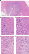
International consensus guidelines for scoring the histopathological growth patterns of liver metastasis
- PMID: 28982110
- PMCID: PMC5680474
- DOI: 10.1038/bjc.2017.334
International consensus guidelines for scoring the histopathological growth patterns of liver metastasis
Abstract
Background: Liver metastases present with distinct histopathological growth patterns (HGPs), including the desmoplastic, pushing and replacement HGPs and two rarer HGPs. The HGPs are defined owing to the distinct interface between the cancer cells and the adjacent normal liver parenchyma that is present in each pattern and can be scored from standard haematoxylin-and-eosin-stained (H&E) tissue sections. The current study provides consensus guidelines for scoring these HGPs.
Methods: Guidelines for defining the HGPs were established by a large international team. To assess the validity of these guidelines, 12 independent observers scored a set of 159 liver metastases and interobserver variability was measured. In an independent cohort of 374 patients with colorectal liver metastases (CRCLM), the impact of HGPs on overall survival after hepatectomy was determined.
Results: Good-to-excellent correlations (intraclass correlation coefficient >0.5) with the gold standard were obtained for the assessment of the replacement HGP and desmoplastic HGP. Overall survival was significantly superior in the desmoplastic HGP subgroup compared with the replacement or pushing HGP subgroup (P=0.006).
Conclusions: The current guidelines allow for reproducible determination of liver metastasis HGPs. As HGPs impact overall survival after surgery for CRCLM, they may serve as a novel biomarker for individualised therapies.
Conflict of interest statement
The authors declare no conflict of interest.
Figures


References
-
- Allison KH, Fligner CL, Parks WT (2004) Radiographically occult, diffuse intrasinusoidal hepatic metastases from primary breast carcinomas: a clinicopathologic study of 3 autopsy cases. Arch Pathol Lab Med 128(12): 1418–1423. - PubMed
-
- Barsky SH, Doberneck SA, Sternlicht MD, Grossman DA, Love SM (1997) 'Revertant' DCIS in human axillary breast carcinoma metastases. J Pathol 183(2): 188–194. - PubMed
-
- Bridgeman VL, Vermeulen PB, Foo S, Bilecz A, Daley F, Kostaras E, Nathan MR, Wan E, Frentzas S, Schweiger T, Hegedus B, Hoetzenecker K, Renyi-Vamos F, Kuczynski EA, Vasudev NS, Larkin J, Gore M, Dvorak HF, Paku S, Kerbel RS, Dome B, Reynolds AR (2017) Vessel co-option is common in human lung metastases and mediates resistance to anti-angiogenic therapy in preclinical lung metastasis models. J Pathol 241(3): 362–374. - PMC - PubMed
-
- Bugyik E, Dezso K, Reiniger L, Laszlo V, Tovari J, Timar J, Nagy P, Klepetko W, Dome B, Paku S (2011) Lack of angiogenesis in experimental brain metastases. J Neuropathol Exp Neurol 70(11): 979–991. - PubMed
Publication types
MeSH terms
LinkOut - more resources
Full Text Sources
Other Literature Sources
Medical
Research Materials

