. 2017 Oct 5;12(10):e0185780.
doi: 10.1371/journal.pone.0185780. eCollection 2017.

NIMA-related kinase 1 (NEK1) regulates meiosis I spindle assembly by altering the balance between α-Adducin and Myosin X

Affiliations

NIMA-related kinase 1 (NEK1) regulates meiosis I spindle assembly by altering the balance between α-Adducin and Myosin X

Miguel A Brieño-Enríquez et al. PLoS One. .

Abstract

NIMA-related kinase 1 (NEK1) is a serine/threonine and tyrosine kinase that is highly expressed in mammalian germ cells. Mutations in Nek1 induce anemia, polycystic kidney and infertility. In this study we evaluated the role of NEK1 in meiotic spindle formation in both male and female gametes. Our results show that the lack of NEK1 provokes an abnormal organization of the meiosis I spindle characterized by elongated and/or multipolar spindles, and abnormal chromosome congression. The aberrant spindle structure is concomitant with the disruption in localization and protein levels of myosin X (MYO10) and α-adducin (ADD1), both of which are implicated in the regulation of spindle formation during mitosis. Interaction of ADD1 with MYO10 is dependent on phosphorylation, whereby phosphorylation of ADD1 enables its binding to MYO10 on mitotic spindles. Reduction in ADD1 protein in NEK1 mutant mice is associated with hyperphosphorylation of ADD1, thereby preventing the interaction with MYO10 during meiotic spindle formation. Our results reveal a novel regulatory role for NEK1 in the regulation of spindle architecture and function during meiosis.


Conflict of interest statement

Competing Interests: The authors have declared that no competing interest exist.

Figures

Fig 1
Fig 1. Loss of NEK1 results in disrupted spindle morphology and chromosome congression during meiosis I in spermatocytes.
(a-c) Immunofluorescence (IF) against β-tubulin (green) and DNA staining with DAPI (magenta) showing meiotic spindle morphology (panels a1-c1), with graphic representations below (panels a2-c2). (A) Nek1+/+ spermatocyte shows normal spindle morphology and chromosome congression at the midplate of the spindle; (b, c) Examples of aberrant spindle morphology in spermatocytes from Nek1kat2j/kat2j mice showing monopolar spindles with mislocalized chromosomes resulting from failed chromosome congression (arrowheads) in b and multipolar spindles in c (arrowheads). (d) Quantitation of abnormal spindles in spermatocytes from Nek1+/+ and Nek1kat2j/kat2j mice (n = 100 cells counted for each). (e) Quantitation cells with abnormal spindles by type of defect. Values are percentages ± Standard deviation. * Indicates statistically significant differences (Unpaired t-test p < 0.05).
Fig 2
Fig 2. Loss of NEK1 results in disrupted spindle morphology and chromosome congression during meiosis I in oocytes.
(a-d) Immunofluorescence (IF) against β-tubulin (green) and DNA staining with DAPI (magenta) showing meiotic spindle morphology (panels a1-d1), with graphic representations below (panels a2-d2). (a) Nek1+/+ oocyte shows normal spindle morphology and chromosome congression at the midplate of the spindle; (b-d) Examples of aberrant spindle morphology in oocytes from Nek1kat2j/kat2j mice showing multipolar spindles with or without errors in chromosome congression (arrowheads indicate three extra spindles formed in the oocyte in b and two additional spindles in c). (e) Quantitation of abnormal spindles in oocytes from Nek1+/+ and Nek1kat2j/kat2j mice (n = 100 cells counted for each). (f) Quantitation of cells with abnormal spindles by type of defect. Values are percentages ± Standard deviation. * Indicates statistically significant differences (Unpaired t-test p < 0.05).
Fig 3
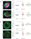
Fig 3. Lack of NEK1 results in abnormal meiosis I spindle morphology in spermatocytes and oocytes.
(a) Immunofluorescence (IF) against β-tubulin (green) and DNA staining with DAPI (magenta) showing a wildtype (top) and Nek1kat2j/kat2j spermatocytes (bottom), along with measurements of spindle length (b) and width (c). Black bars are means (μm) ± Standard deviation. (d) Immunofluorescence (IF) against β-tubulin (green) and DNA staining with DAPI (magenta) showing a wildtype (top) and Nek1kat2j/kat2j (bottom) oocytes, along with measurements of spindle length (b) and width (c). Black bars are means (μm) ± Standard deviation. * Indicates statistically significant differences (Unpaired t-test p < 0.05).
Fig 4
Fig 4. Nek1kat2j/kat2j spermatocytes and oocytes show aberrant localization of myosin X (MYO10) on meiosis I spindles.
(a) IF against MYO10 (red), β-tubulin (green) and DAPI (magenta) on spermatocytes from wildtype (left) and Nek1kat2j/kat2j mice (right) in coronal (top) and transverse (bottom) planes. Arrowheads indicate examples of MYO10 foci. Quantitation of MYO10 focus counts per nucleus are given in panel (b). (c) IF against MYO10 (red), β-tubulin (green) and DAPI (magenta) in oocytes of both wildtype and mutant mice. Arrowheads indicate examples of MYO10 foci. Quantitation of MYO10 focus counts per nucleus are given in panel (d). All values are focus counts per spindle and black bars denote means ± Standard deviation. * Indicates statistically significant differences (Unpaired t test p < 0.05).
Fig 5
Fig 5. Nek1kat2j/kat2j spermatocytes and oocytes show aberrant localization of Adducin 1 (ADD1) on meiosis I spindles.
(a) IF against ADD1 (red), β-tubulin (green) and DAPI (magenta) on spermatocytes from wildtype (left) and Nek1kat2j/kat2j mice (right) in coronal (top) and transverse (bottom) planes. Arrowheads indicate examples of ADD1 foci. Quantitation of ADD1 focus counts per nucleus are given in panel (b). (c) IF against ADD1 (red), β-tubulin (green) and DAPI (magenta) in oocytes of both wildtype and mutant mice. Arrowheads indicate examples of ADD1 foci. Quantitation of ADD1 focus counts per nucleus are given in panel (d). All values are focus counts per spindle and black bars denote means ± Standard deviation. * Indicates statistically significant differences (Unpaired t test p < 0.05).
Fig 6
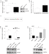
Fig 6. Loss of NEK1 during meiosis is associated with hyper-phosphorylation and reduced ADD1 protein in a PP1γ-dependent manner.
(a) Quantitation of ADD1 protein levels in Nek1+/+ testis extracts relative to GAPDH control. (b) ADD1 and MYO10 protein levels, expressed as a ratio of Nek1kat2j/kat2j / Nek1+/+, as determined by mass spectrometry. (c) ADD1 phosphopeptide and deaminated peptide sequence determined by mass spectrometry to be higher in Nek1kat2j/kat2j testis extracts. (d) Quantitation by western blotting of ADD1 protein levels in oocytes at 0h and after 6h of culture. (e) Quantification of ADD1 protein levels in wildtype oocytes cultured for 6h in vehicle (ethanol) and the PP1 inhibitor Calyculin A (CLA) (at doses of 2 and 20 nM). All values are means ± Standard deviation. * Indicate statistically significant differences (Unpaired t test p < 0.05 and one-way ANOVA followed by Dunnett’s multiple comparisons test, p < 0.05, for cultures with inhibitor)

References

    1. Gray S, Cohen PE. Control of Meiotic Crossovers: From Double-Strand Break Formation to Designation. Annu Rev Genet 2016;50: 175–210. doi: 10.1146/annurev-genet-120215-035111 - DOI - PMC - PubMed
    1. Duro E, Marston AL. From equator to pole: splitting chromosomes in mitosis and meiosis. Genes Dev 2015;29: 109–122. doi: 10.1101/gad.255554.114 - DOI - PMC - PubMed
    1. Bennabi I, Terret ME, Verlhac MH. Meiotic spindle assembly and chromosome segregation in oocytes. J Cell Biol 2016;215: 611–619. doi: 10.1083/jcb.201607062 - DOI - PMC - PubMed
    1. Ohkura H. Meiosis: an overview of key differences from mitosis. Cold Spring Harb Perspect Biol 2015;7. - PMC - PubMed
    1. McKim KS, Hawley RS. Chromosomal control of meiotic cell division. Science 1995;270: 1595–1601. - PubMed