. 2017 Oct 5;36(1):138.
doi: 10.1186/s13046-017-0604-3.

MiR-203a-3p suppresses cell proliferation and metastasis through inhibiting LASP1 in nasopharyngeal carcinoma

Affiliations

MiR-203a-3p suppresses cell proliferation and metastasis through inhibiting LASP1 in nasopharyngeal carcinoma

Ning Jiang et al. J Exp Clin Cancer Res. .

Abstract

Background: miR-203a-3p was reported as a tumor suppressor and disregulated in many malignancies including nasopharyngeal carcinoma (NPC). However, its function in tumor growth and metastasis in NPC has rarely been reported.

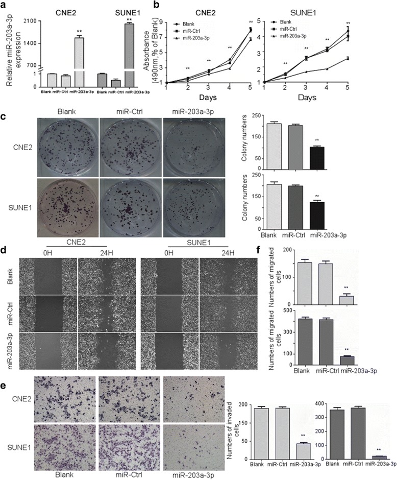
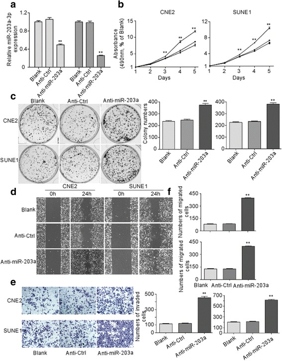
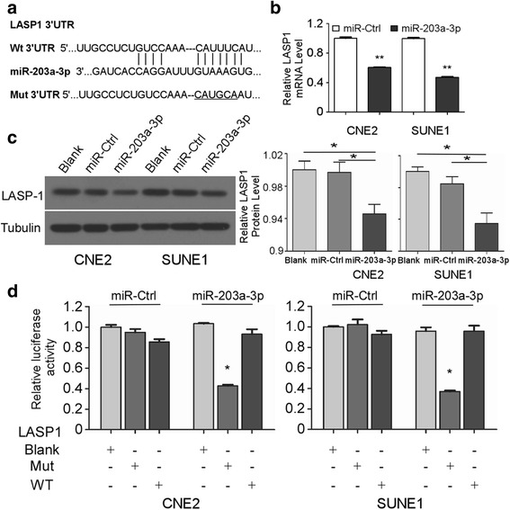
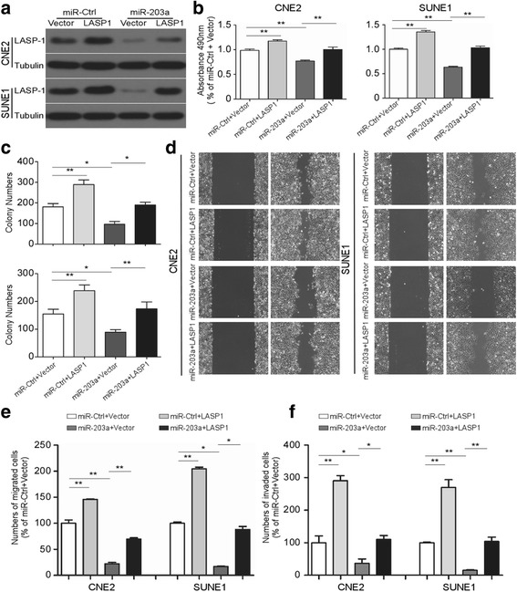
Methods: The expression level of miR-203a-3p in human NPC tissues and cell lines was detected via real-time PCR (RT-PCR). Cell proliferation, migration and invasion were assessed in vitro by MTT, colony formation and transwell assay, respectively. The function of miR-203a-3p in vivo was detected through NPC xenograft tumor growth and lung metastatic mice model. Dual-luciferase reporter assay was used to identify the direct target of miR-203a-3p.

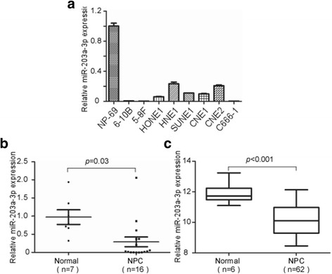
Results: The expression of miR-203a-3p was decreased in NPC tissues and cell lines in comparison with normal nasopharyngeal tissues and cell line. Ectopic expression of miR-203a-3p inhibited while inhibiting miR-203a-3p expression increased NPC cell proliferation, migration and invasion in vitro. MR-203a-3p overexpression suppressed xenograft tumor growth and lung metastasis in vivo. LASP1 was identified as a direct target of miR-203a-3p, which was confirmed by real-time PCR and western blotting assay. Ectopic expression of LASP1 partially reversed miR-203a-3p-mediated inhibition on proliferation, migration and invasion in NPC cells.

Conclusion: Collectively, miR-203a-3p suppresses tumor growth and metastasis through targeting LASP1 in NPC. The newly identified miR-203a-3p/LASP1 pathway provides further insights into the initiation and progression of NPC, which may represent a novel therapeutic target for NPC.

Keywords: LASP1; Nasopharyngeal carcinoma; Proliferation; metastasis; miR-203a-3p.


Conflict of interest statement

Ethics approval and consent to participate

All procedures performed in studies involving human participants were in accordance with the ethical standards of the Ethics Committee of the Institutional Ethical Review Board of Jiangsu Cancer Hospital. All patients studied signed an informed consent for participation. All animal procedures and care were conducted in accordance with institutional guidelines and in compliance with national and international laws and policies.

Competing interests

The authors declare that they have no competing interests.

Publisher’s Note

Springer Nature remains neutral with regard to jurisdictional claims in published maps and institutional affiliations.

Figures

Fig. 1
Fig. 1
MiR-203a-3p is downregulated in NPC cell lines and clinical specimens. Relative expression of miR-203a-3p in NP69 and NPC cell lines by real-time PCR a. Relative expression of miR-203a-3p in normal nasopharyngeal epithelial (n = 7) and NPC tissues (n = 16) b. Each experiment was independently repeated for at least three times. Data are presented as mean ± S.D. MiR-203a-3p expression profile in 62 NPC and 6 normal nasopharyngeal epithelial samples using GEO database c. P-value was calculated using the Student’s t-test
Fig. 2
Fig. 2
Ectopic expression of MiR-203a-3p suppresses NPC cell proliferation, invasion and migration in vitro. CNE2 and SUNE1 cells were transfected with miR-203a-3p mimic (50 nM), miR-Ctrl (50 nM), or the same volume of PBS (Blank). Expression of miR-203a-3p after transfection a. MTT assays were performed in CNE2 and SUNE1 cells on one to five days after transfection b. Colony formation was performed by crystal violet staining in CNE2 and SUNE1 cells c. Representative images for wound healing assay d and transwell invasion assay e. The cell counting results of transwell migration f and invasion assay e. * P < 0.05; ** P < 0.01 compared with miR-Ctrl or Blank groups. Each experiment was independently repeated at least three times. Data are presented as mean ± S.D. Statistical analysis was performed using one way ANOVA
Fig. 3
Fig. 3
MiR-203a-3p inhibition increased NPC cell proliferation, invasion and migration in vitro. CNE2 and SUNE1 cells were transfected with miR-203a-3p inhibitor (Anti-miR-203a, 100 nM), negative control (Anti-Ctrl, 100 nM) or PBS (Blank). Expression of miR-203a-3p after transfection a. MTT assays were performed in CNE2 and SUNE1 cells on one to five days after transfection b. Colony formation was performed by crystal violet staining in CNE2 and SUNE1 cells c. Representative images for wound healing assay d and transwell invasion assays e. The cell counting results of transwell migration e and invasion assays f. ** P < 0.01 compared with Anti-Ctrl or Blank groups. The results are representative of at least three independent experiments. Statistical analysis was performed using one way ANOVA
Fig. 4
Fig. 4
MiR-203a-3p inhibits tumor growth and metastasis in NPC in vivo . SUNE1 cells stably overexpressing miR-203a (Lenti-miR-203a) or negative control empty pSin-EF2-vector (Lenti-vector) were subcutaneously injected into right flank of each nude mouse (n = 6). A photograph of nude mice carrying tumors a. Volumes of all tumors were detected every 3 days b. SUNE-1 cells stably overexpressing miR-203a (Lenti-miR-203a) or negative control empty lenti-vector (Lenti-vector) were intravenously injected via the tail vein and the formation of lung metastases was assessed after 8 weeks. Representative images c and quantification d of macroscopic metastatic nodules on the lung surface. Representative images e and quantification f of microscopic metastatic nodules in lung tissue sections stained with hematoxylin and eosin (×100). Data are presented as mean ± S.D.; ** P < 0.01 compared with the Lenti-vector group, Student’s t-test
Fig. 5
Fig. 5
LASP1 is a direct target of miR-203a-3p in NPC cells. Wt or Mt. of the LASP1 mRNA 3′-UTR sequences targeted by miR-203a-3p a. Quantification of LASP1 mRNA levels by quantitative RT-PCR b and LASP1 protein expression by western blotting c after transfection with miR-203a-3p mimic or miR-Ctrl. Relative luciferase activity of CNE-2 and SUNE-1 cells after co-transfection with Wt or Mt. LASP1 3′-UTR reporter genes (2 μg) and miR-203a-3p mimic or miR-Ctrl (50 nM) d. Each experiment was independently repeated at least three times. Data are presented as mean ± S.D.; * P < 0.05; ** P < 0.01 compared with the miR-Ctrl group, Student’s t-test
Fig. 6
Fig. 6
LASP1 is involved in miR-203a-3p mediated tumor suppression. CNE-2 and SUNE-1 cells were co-transfected with either miR-203a-3p mimic or negative control (miR-203a or miR-Ctrl) and either the pSin-EF2-puro-LASP1 (LASP1) plasmid or empty pSin-EF2-puro-Vector (Vector). a Western blotting analysis of LASP1 protein expression. b-f Overexpression of LASP1 abrogated the inhibition on cell proliferation, migration and invasion by miR-203a-3p. Representative results of the MTT assay (b), colony formation assay (c), wounding healing assay (d), transwell migration assay (e) and invasion assay (f). Data are presented as mean ± S.D.; * P < 0.05, ** P < 0.01, one way ANOVA. Each experiment was independently repeated at least three times

References

    1. McDermott AL, Dutt SN, Watkinson JC. The aetiology of nasopharyngeal carcinoma. Clin Otolaryngol Allied Sci. 2001;26:82–92. doi: 10.1046/j.1365-2273.2001.00449.x. - DOI - PubMed
    1. Chen W, Zheng R, Baade PD, Zhang S, Zeng H, Bray F, et al. Cancer statistics in China, 2015. CA Cancer J Clin. 2016;66:115–132. doi: 10.3322/caac.21338. - DOI - PubMed
    1. Lai SZ, Li WF, Chen L, Luo W, Chen YY, Liu LZ, et al. How does intensity-modulated radiotherapy versus conventional two-dimensional radiotherapy influence the treatment results in nasopharyngeal carcinoma patients? Int J Radiat Oncol Biol Phys. 2011;80:661–668. doi: 10.1016/j.ijrobp.2010.03.024. - DOI - PubMed
    1. Cheng SK, Dizon J. Computerised cognitive behavioural therapy for insomnia: a systematic review and meta-analysis. Psychother Psychosom. 2012;81:206–216. doi: 10.1159/000335379. - DOI - PubMed
    1. Guo Q, Pan J, Zong J, Zheng W, Zhang C, Tang L, et al. Suggestions for lymph node classification of UICC/AJCC staging system: a retrospective study based on 1197 nasopharyngeal carcinoma patients treated with intensity-modulated radiation therapy. Medicine (Baltimore) 2015;94:e808. doi: 10.1097/MD.0000000000000808. - DOI - PMC - PubMed

LinkOut - more resources