. 2017 Oct 5;17(1):26.
doi: 10.1186/s40644-017-0128-2.

Lymphomatosis cerebri: a rare variant of primary central nervous system lymphoma and MR imaging features

Affiliations

Lymphomatosis cerebri: a rare variant of primary central nervous system lymphoma and MR imaging features

Hui Yu et al. Cancer Imaging. .

Abstract

Background: Lymphomatosis cerebri (LC) is a rare variant of primary central nervous system lymphoma (PCNSL), characterized by diffuse infiltration without the formation of a discrete mass. The diagnosis of LC is a challenge because the imaging findings are atypical for lymphoma. The purpose of present study is to investigate MRI characteristics and clinical features of LC and potentially facilitate an early and accurate diagnosis of this often-missed disease.

Methods: Seven patients (average 44 years, 19-58 years) with LC proved basing on MRI and histology were retrospectively reviewed the clinical data and cerebral MR imaging findings.

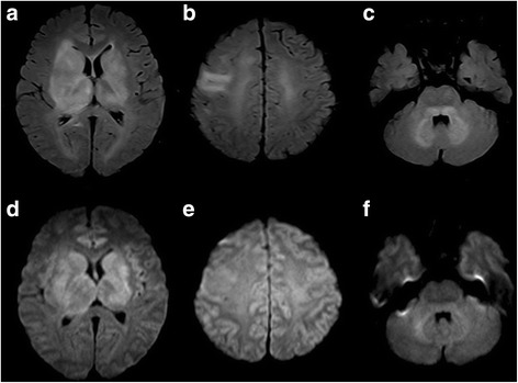
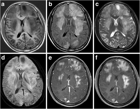
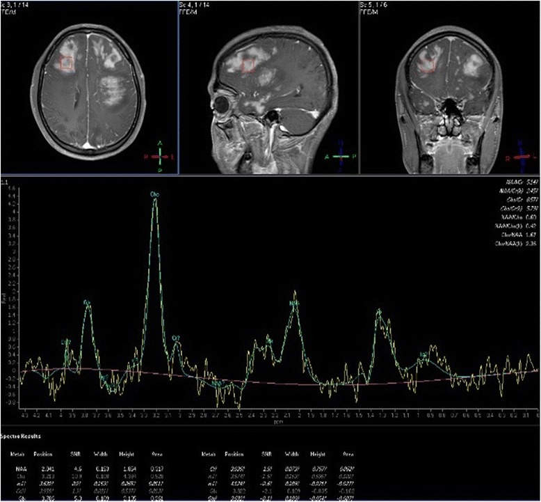
Results: The common presenting symptoms were cognitive decline, behavioral disturbance, gait disturbance. All patients had both deep and lobar lesion distribution, and two of them had infratentorial involvement. Lack of contrast enhancement and subtle patchy enhanced pattern were observed in two and three patients, respectively. The remaining two patients presented multiple patchy enhancement. Most of the lesions were slightly hyperintense to normal brain on DWI as well as hyperintense on ADC maps. Three patients presented a pattern of marked decrease of NAA/Cr, increase of Cho/Cr, and two of the three cases showed increased Lip/Cr and Lac/Cr on MRS.

Conclusions: We conclude that diffuse bilateral lesions especially in deep and lobar region including white and gray matter, without enhancement or with patchy enhancement, marked decrease of NAA/Cr and increase of Cho/Cr, and increased Lip/Cr and Lac/Cr are suggestive of LC. Prompt recognition of these imaging patterns may lead to early diagnosis of LC and brain biopsy with improved prognosis.

Keywords: Diffusion weighted imaging; Lymphomatosis cerebri; Magnetic resonance imaging; Magnetic resonance spectroscopy; Primary central nervous system lymphoma.


Conflict of interest statement

Ethics approval and consent to participate

All patients provided an informed consent for MRI examination and for the use of personal data. Therefore, for this retrospective analysis ethics committee approval was not requested. All data, including images, were anonymized and all patients provided their consent for the use of personal data.

Consent for publication

All the patients have given a written consent to use their data for research purpose, including publications in medical journals.

Competing interests

The authors declare that they have no competing interests.

Publisher’s Note

Springer Nature remains neutral with regard to jurisdictional claims in published maps and institutional affiliations.

Figures

Fig. 1
Fig. 1
Nineteen-year-old man with lymphomatosis cerebri. a, b and c, Axial T2-weighted-FLAIR with STIR show the lesions distribution of type I, II and III respectively, which involved bilateral superior cerebellar peduncles, dentate nucleus of cerebellum, basal ganglia, internal capsule, thalamus and right frontal lobes. d, e and f, DWI images show slight hyperintensity in the lesions, but corresponding ADC maps (not shown) indicate no water of restriction
Fig. 2
Fig. 2
Forty five-year-old woman with lymphomatosis cerebri. a, b and c, Axial T1WI, T2WI and T2-weighted-FLAIR show the distribution of type I and II lesions, which involved bilateral frontal lobes and basal ganglia, left thalamus, and genu of corpus callosum. d and e , DWI shows slight hyperintensityand corresponding low ADC in portions of the genu of corpus callosum and left frontal lobe. f , Post-contrast T1WI shows subtle patchy contrast enhancement in the left frontal lobe
Fig. 3
Fig. 3
Twenty one-year-old man with lymphomatosis cerebri. a, b and c, Axial T1WI,T2-weighted FLAIR and T2WI show the distribution of type I and II lesions, which involved bilateral frontal lobes, occipital lobes, basal ganglia and insula. d DWI shows slight hyperintensity in the lesions, which were also slightly hyperintense on ADC map(not shown). e Post-contrast T1WI shows multiple patchy contrast enhancement in the lesions. f, Delayed scanning of post-contrast T1WI shows extended enhancement range of the lesions
Fig. 4
Fig. 4
The same case shown in Fig.2. Multiple-voxel spectra are acquired with a point-resolved spectroscopy (PRESS) sequence, TR 1500 ms, TE 135 ms, NSA 128. MRS shows elevation of Cho/Cr and marked reduction of NAA/Cr in the lesion. In addition, there is an inverted Lac peak
Fig. 5
Fig. 5
The same case shown in Fig.3. Single-voxel spectra are acquired with a point-resolved spectroscopy (PRESS) sequence, TR 2000 ms, TE 40 ms, NSA 128. MRS shows elevation of Cho/Cr and marked reduction of NAA/Cr in the lesion. In addition, there is a large Lip peak
Fig. 6
Fig. 6
a Brain biopsy specimen from the case 1 shows dispersed round neoplastic cells spreading along the white matter tracts without causing tissue destruction or mass formation and are occasionally clustered around blood vessels (H&E × 400). b Immunohistochemistry shows that the atypical cells were positive for CD20, a marker for b cells

References

    1. Hatanpaa KJ, Fuda F, Koduru P, Young K, Lega B, Chen W. Lymphomatosis Cerebri: A Diagnostic Challenge. JAMA Neurol. 2015;72:1066–1067. doi: 10.1001/jamaneurol.2015.1149. - DOI - PubMed
    1. Bakshi R, Mazziotta JC, Mischel PS, Jahan R, Seligson DB, Vinters HV. Lymphomatosis cerebri presenting as a rapidly progressive dementia: clinical, neuroimaging and pathologic findings. Dement Geriatr Cogn Disord. 1999;10:152–157. doi: 10.1159/000017116. - DOI - PubMed
    1. Kitai R, Hashimoto N, Yamate K, Ikawa M, Yoneda M, Nakajima T, Arishima H, Takeuchi H, Sato K, Kikuta K. Lymphomatosis cerebri: clinical characteristics, neuroimaging, and pathological findings. Brain Tumor Pathol. 2012;29:47–53. doi: 10.1007/s10014-011-0064-y. - DOI - PubMed
    1. Lewerenz J, Ding XQ, Matschke J, Schnabel C, Emami P, von Borczyskowski D, Buchert R, Krieger T, de Wit M, Munchau A. Dementia and leukoencephalopathy due to lymphomatosis cerebri. BMJ Case Rep. 2009;2009 10.1136/bcr.08.2008.0752. - PMC - PubMed
    1. Izquierdo C, Velasco R, Vidal N, Sanchez JJ, Argyriou AA, Besora S, Graus F, Bruna J. Lymphomatosis cerebri: a rare form of primary central nervous system lymphoma. Analysis of 7 cases and systematic review of the literature. Neuro-Oncology. 2016;18:707–715. doi: 10.1093/neuonc/nov197. - DOI - PMC - PubMed