

MR/PET Imaging of the Cardiovascular System
- PMID: 28982570
- PMCID: PMC6415529
- DOI: 10.1016/j.jcmg.2017.07.008
MR/PET Imaging of the Cardiovascular System
Abstract
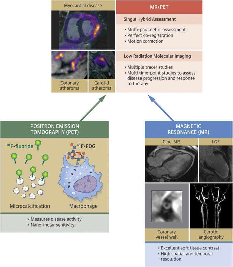
Cardiovascular imaging has largely focused on identifying structural, functional, and metabolic changes in the heart. The ability to reliably assess disease activity would have major potential clinical advantages, including the identification of early disease, differentiating active from stable conditions, and monitoring disease progression or response to therapy. Positron emission tomography (PET) imaging now allows such assessments of disease activity to be acquired in the heart, whereas magnetic resonance (MR) scanning provides detailed anatomic imaging and tissue characterization. Hybrid MR/PET scanners therefore combine the strengths of 2 already powerful imaging modalities. Simultaneous acquisition of the 2 scans also provides added benefits, including improved scanning efficiency, motion correction, and partial volume correction. Radiation exposure is lower than with hybrid PET/computed tomography scanning, which might be particularly beneficial in younger patients who may need repeated scans. The present review discusses the expanding clinical literature investigating MR/PET imaging, highlights its advantages and limitations, and explores future potential applications.
Keywords: MR; PET; atherosclerosis; cardiomyopathy; hybrid imaging.
Copyright © 2017 The Authors. Published by Elsevier Inc. All rights reserved.
Figures
Similar articles
-
Applications of PET-MR Imaging in Cardiovascular Disorders.PET Clin. 2020 Oct;15(4):509-520. doi: 10.1016/j.cpet.2020.06.007. Epub 2020 Jul 21. PET Clin. 2020. PMID: 32888548 Free PMC article. Review.
-
Coronary Artery PET/MR Imaging: Feasibility, Limitations, and Solutions.JACC Cardiovasc Imaging. 2017 Oct;10(10 Pt A):1103-1112. doi: 10.1016/j.jcmg.2016.09.029. Epub 2017 Jan 18. JACC Cardiovasc Imaging. 2017. PMID: 28109921 Free PMC article.
-
Hybrid positron emission tomography-magnetic resonance of the heart: current state of the art and future applications.Eur Heart J Cardiovasc Imaging. 2018 Sep 1;19(9):962-974. doi: 10.1093/ehjci/jey090. Eur Heart J Cardiovasc Imaging. 2018. PMID: 30010838 Free PMC article. Review.
-
Cardiac Applications of PET/MR Imaging.Magn Reson Imaging Clin N Am. 2017 May;25(2):325-333. doi: 10.1016/j.mric.2016.12.007. Epub 2017 Feb 27. Magn Reson Imaging Clin N Am. 2017. PMID: 28390532 Review.
-
PET/MR imaging: technical aspects and potential clinical applications.Radiology. 2013 Apr;267(1):26-44. doi: 10.1148/radiol.13121038. Radiology. 2013. PMID: 23525716 Review.
Cited by
-
Applications of PET-MR Imaging in Cardiovascular Disorders.PET Clin. 2020 Oct;15(4):509-520. doi: 10.1016/j.cpet.2020.06.007. Epub 2020 Jul 21. PET Clin. 2020. PMID: 32888548 Free PMC article. Review.
-
Hybrid PET/MRI of large vessel vasculitis : Radiation dose compared to PET/CT with view on cumulative effective dose.Wien Klin Wochenschr. 2024 Nov;136(21-22):627-635. doi: 10.1007/s00508-024-02336-2. Epub 2024 Mar 8. Wien Klin Wochenschr. 2024. PMID: 38456940 Free PMC article.
-
18F-FDG-PET/MR imaging to monitor disease activity in large vessel vasculitis.Nat Commun. 2024 Aug 25;15(1):7314. doi: 10.1038/s41467-024-51613-1. Nat Commun. 2024. PMID: 39183340 Free PMC article.
-
Scan-rescan measurement repeatability of 18F-FDG PET/MR imaging of vascular inflammation.J Nucl Cardiol. 2022 Aug;29(4):1660-1670. doi: 10.1007/s12350-021-02627-5. Epub 2021 May 27. J Nucl Cardiol. 2022. PMID: 34046803
-
Cardiovascular 18F-fluoride positron emission tomography-magnetic resonance imaging: A comparison study.J Nucl Cardiol. 2021 Oct;28(5):1-12. doi: 10.1007/s12350-019-01962-y. Epub 2019 Dec 2. J Nucl Cardiol. 2021. PMID: 31792913 Free PMC article.
References
-
- Sarikaya I Cardiac applications of PET. Nucl Med Commun 2015;36:971–85. - PubMed
-
- Bergmann SR, Weinheimer CJ, Markham J, Herrero P. Quantitation of myocardial fatty acid metabolism using PET. J Nucl Med 1996;37: 1723–30. - PubMed
-
- Tillisch J, Brunken R, Marshall R, et al. Reversibility of cardiac wall-motion abnormalities predicted by positron tomography. N Engl J Med 1986;314:884–8. - PubMed
-
- Ohira H, Tsujino I, Ishimaru S, et al. Myocardial imaging with 18F-fluoro-2-deoxyglucose positron emission tomography and magnetic resonance imaging in sarcoidosis. Eur J Nucl Med Mol Imaging 2008;35:933–41. - PubMed
-
- Youssef G, Leung E, Mylonas I, et al. The use of 18F-FDG PET in the diagnosis of cardiac sarcoidosis: a systematic review and metaanalysis including the Ontario experience. J Nucl Med 2012;53:241–8. - PubMed
Publication types
MeSH terms
Substances
Grants and funding
LinkOut - more resources
Full Text Sources
Other Literature Sources
Medical
