Review
. 2017 Oct;10(10 Pt A):1180-1193.
doi: 10.1016/j.jcmg.2017.08.005.

Role of Cardiac Magnetic Resonance in the Diagnosis and Prognosis of Nonischemic Cardiomyopathy

Affiliations
Review

Role of Cardiac Magnetic Resonance in the Diagnosis and Prognosis of Nonischemic Cardiomyopathy

Amit R Patel et al. JACC Cardiovasc Imaging. 2017 Oct.

Abstract

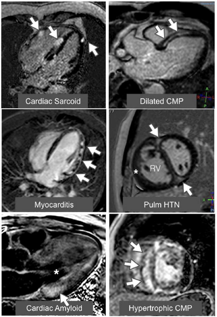
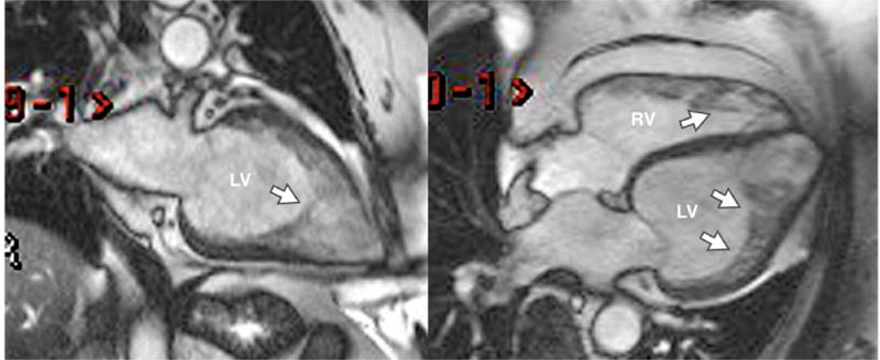
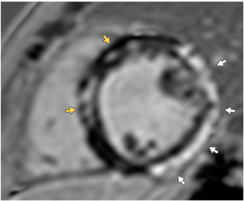
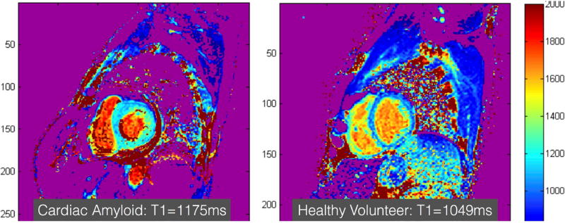
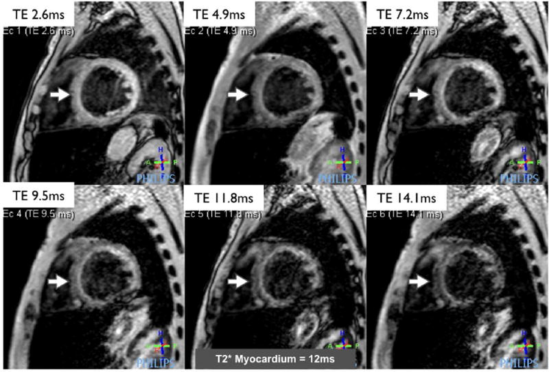
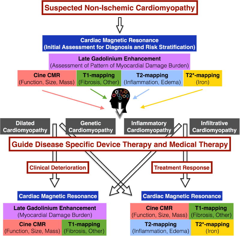
Cardiac magnetic resonance (CMR) is a valuable tool for the evaluation of patients with, or at risk for, heart failure and has a growing impact on diagnosis, clinical management, and decision making. Through its ability to characterize the myocardium by using multiple different imaging parameters, it provides insight into the etiology of the underlying heart failure and its prognosis. CMR is widely accepted as the reference standard for quantifying chamber size and ejection fraction. Additionally, tissue characterization techniques such as late gadolinium enhancement (LGE) and other quantitative parameters such as T1 mapping, both native and with measurement of extracellular volume fraction; T2 mapping; and T2* mapping have been validated against histological findings in a wide range of clinical scenarios. In particular, the pattern of LGE in the myocardium can help determine the underlying etiology of the heart failure. The presence and extent of LGE determine prognosis in many of the nonischemic cardiomyopathies. The use of CMR should increase as its utility in characterization and assessment of prognosis in cardiomyopathies is increasingly recognized.

Keywords: amyloidosis; cardiomyopathy; cardiovascular magnetic resonance; heart failure; hypertrophic cardiomyopathy; sarcoidosis.


Figures

Figure 1
Figure 1. Examples of late gadolinium enhancement (LGE) in a variety of nonischemic cardiomyopathies
The top left image shows a 4-chamber view of a patchy distribution of late mid wall and epicardial LGE in a patient with cardiac sarcoidosis. The top right image shows a 3-chamber view of a mid-wall stripe pattern of LGE in a patient with dilated cardiomyopathy. The middle left image shows a 4-chamber view of patchy epicardial and midwall LGE along the lateral wall in a patient with myocarditis. The middle right image shows a mid-ventricular short axis image in a patient with pulmonary hypertension with right ventricular (RV) dilation and hypertrophy (*) along with LGE in the anterior and inferior right ventricular insertion points (arrows). The bottom left image shows a 3-chamber view of a LGE image in an amyloid patient. The left ventricular blood pool is nulled (*) and there is subtle circumferential subendocardial LGE throughout the left ventricle (LV). The LGE is most pronounced at the base of LV within hypertrophied myocardium. The bottom right image shows a mid-ventricular short axis image in a patient with HCM with evidence of asymmetric septal hypertrophy with extensive mid-wall LGE within the hypertrophied myocardium.
Figure 2
Figure 2. LV Noncompaction
Diastolic still frames from cine images of a two chamber view (left) and four chamber view (right) are shown. The myocardium is thin and the left ventricle (LV) and right ventricle (RV) are heavily trabeculated (arrows).
Figure 3
Figure 3. Duchenne’s Muscular Dystrophy
A short axis image of the mid ventricle obtained in a patient with Duchenne’s Muscular Dystrophy shows epicardial late gadolinium enhancement (LGE) along the lateral wall (white arrows) and mid-wall LGE in the septum.
Figure 4
Figure 4. Cardiac amyloidosis
Short axis images of the mid ventricle encoded with a native myocardial T1-color map generated from a series of images acquired at increasing repetition times acquired using MOdified Look Locker Image (MOLLI) pulse sequence is shown in a patient with cardiac amyloidosis (left) and a healthy volunteer (right). The T1-time shown in this figure are obtained from a region of interest drawn manually in the inter-ventricular septum. The T1-times can be even higher in patients with more extensive cardiac amyloidosis than the example shown here. Normal native T1 value on the particular scanner used to acquire these images is <1050ms.
Figure 5
Figure 5. Cardiac siderosis
A series of left ventricular short axis images acquired using a single breath-hold T2* pulse sequence in a patient with sickle cell anemia. A series of images are acquired at increasing echo times (TE). The signal intensity in the myocardium decreases as the TE increases. The rate of decay is used to calculate the T2* relaxation time, which is reduced in this patient and indicates the presence of significant myocardial iron overload.
Central Illustration
Central Illustration. Evaluation of Non-ischemic Cardiomyopathy Using Cardiac Magnetic Resonance
This chart demonstrates a potential approach for incorporating the use of cardiac magnetic resonance for the initial evaluation and follow up of patients with cardiomyopathy.

Similar articles

Cited by

References

    1. Abbasi SA, Ertel A, Shah RV, et al. Impact of cardiovascular magnetic resonance on management and clinical decision-making in heart failure patients. Journal of cardiovascular magnetic resonance : official journal of the Society for Cardiovascular Magnetic Resonance. 2013;15:89. - PMC - PubMed
    1. Kramer CM. Role of Cardiac MR Imaging in Cardiomyopathies. Journal of nuclear medicine : official publication, Society of Nuclear Medicine. 2015;56(Suppl 4):39S–45S. - PMC - PubMed
    1. Arbustini E, Narula N, Tavazzi L, et al. The MOGE(S) classification of cardiomyopathy for clinicians. Journal of the American College of Cardiology. 2014;64:304–18. - PubMed
    1. McMurray JJ, Packer M, Desai AS, et al. Angiotensin-neprilysin inhibition versus enalapril in heart failure. The New England journal of medicine. 2014;371:993–1004. - PubMed
    1. Iles LM, Ellims AH, Llewellyn H, et al. Histological validation of cardiac magnetic resonance analysis of regional and diffuse interstitial myocardial fibrosis. European heart journal cardiovascular Imaging. 2015;16:14–22. - PubMed

Publication types

MeSH terms