. 2017 Nov 1;37(44):10725-10737.
doi: 10.1523/JNEUROSCI.1164-17.2017. Epub 2017 Oct 5.

Neural Signature of Value-Based Sensorimotor Prioritization in Humans

Affiliations

Neural Signature of Value-Based Sensorimotor Prioritization in Humans

Annabelle Blangero et al. J Neurosci. .

Abstract

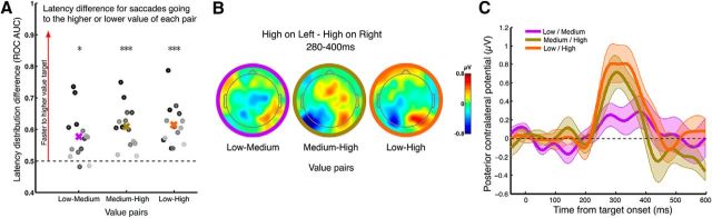
In situations in which impending sensory events demand fast action choices, we must be ready to prioritize higher-value courses of action to avoid missed opportunities. When such a situation first presents itself, stimulus-action contingencies and their relative value must be encoded to establish a value-biased state of preparation for an impending sensorimotor decision. Here, we sought to identify neurophysiological signatures of such processes in the human brain (both female and male). We devised a task requiring fast action choices based on the discrimination of a simple visual cue in which the differently valued sensory alternatives were presented 750-800 ms before as peripheral "targets" that specified the stimulus-action mapping for the upcoming decision. In response to the targets, we identified a discrete, transient, spatially selective signal in the event-related potential (ERP), which scaled with relative value and strongly predicted the degree of behavioral bias in the upcoming decision both across and within subjects. This signal is not compatible with any hitherto known ERP signature of spatial selection and also bears novel distinctions with respect to characterizations of value-sensitive, spatially selective activity found in sensorimotor areas of nonhuman primates. Specifically, a series of follow-up experiments revealed that the signal was reliably invoked regardless of response laterality, response modality, sensory feature, and reward valence. It was absent, however, when the response deadline was relaxed and the strategic need for biasing removed. Therefore, more than passively representing value or salience, the signal appears to play a versatile and active role in adaptive sensorimotor prioritization.SIGNIFICANCE STATEMENT In many situations such as fast-moving sports, we must be ready to act fast in response to sensory events and, in our preparation, prioritize courses of action that lead to greater rewards. Although behavioral effects of value biases in sensorimotor decision making have been widely studied, little is known about the neural processes that set these biases in place beforehand. Here, we report the discovery of a transient, spatially selective neural signal in humans that encodes the relative value of competing decision alternatives and strongly predicts behavioral value biases in decisions made ∼500 ms later. Follow-up manipulations of value differential, reward valence, response modality, sensory features, and time constraints establish that the signal reflects an active, feature- and effector-general preparatory mechanism for value-based prioritization.

Keywords: ERP; decision making; human; sensorimotor; urgency; value bias.


Figures

Figure 1.
Figure 1.
Original value-biased, speeded discrimination task and behavioral signatures of prioritization. A, Trial timeline of the main task. Subjects were presented with two colored discs (“targets”), respectively, associated with five and 40 points. After a delay, the central fixation color changed to one of the two alternatives, instructing the subjects to make an immediate saccade to the corresponding target within a tight deadline to earn the corresponding number of points. B, Saccade RT distributions pooled across subjects showing higher error rates and longer correct RTs for lower-value cues. C, Individual error rate differences (low-value minus high-value cues) plotted against RT effects (ROC AUC for correct high-value versus low-value saccades) revealing a wide range across subjects in the degree of behavioral value-biases.
Figure 2.
Figure 2.
Electrophysiological signature of relative valuation in advance of the imperative visual discrimination in the original task. A, Electrode × time map of t-values quantifying the effect of placement of the higher value target on the left versus right side of the display. To identify spatially selective, value-encoding activity specifically, electrodes were considered in symmetric pairs ordered from anterior to posterior and the minimum absolute t-value across the two hemispheres is marked only for time points/electrode pairs having a signed t-value of opposite polarity on the left versus right hemisphere. This analysis highlighted a PCP, which, as shown in the following sections, is replicated in several other experiments in the same subjects (Fig. 4) and also in a different set of subjects (Fig. 5). B, Topographic map of the differential potential for high-on-left minus high-on-right trials integrated over the time period 280–400 ms, along with the waveforms for the individual conditions from the electrodes at the left (PO7) and right (PO8) focus. C, Average contralateral signal for four subgroups of subjects indicated in Figure 1C.
Figure 3.
Figure 3.
Behavior prediction by the PCP. A, Intersubject correlation between the amplitude of the PCP and the value-based, starting-point bias parameter of the diffusion model. B, Saccade RT distributions for trials with a larger (left) and smaller (right) single-trial PCP amplitude with respect to the median. The inset shows the individual starting point bias parameter fits for large versus small PCP, highlighting the consistency of the relative difference in bias. C, Individual ROC area values for the prediction of error versus correct action on low-value cued trials based on single-trial PCP amplitude. Single-trial measures of PCP amplitude were taken from the discriminating “component” derived from linear classification of high-on-left trials versus high-on-right trials, conditions that, critically, are independent of behavioral outcome. ROC analysis for prediction of correct/incorrect behavioral outcome was then performed within each of these trial types separately. Due to the manner in which PCP amplitude was derived, ROC area values come out >0.5 (chance) for high-on-left trials but <0.5 for high-on-right, indicating that, in both conditions separately, a greater PCP predicts a greater likelihood of erroneous saccade toward the higher value target on a low-value cue.
Figure 4.
Figure 4.
Additional experiments addressing feature and effector general nature of the PCP. Eight subjects who showed strong behavioral biases in the original task also participated in follow-up experiments. A, Data from the original task in just these eight subjects. B, Data from a unilateral button press version of the task. C, Data from a shape-value version of the task. D, Data from a losses (instead of gains) version of the task. Across all panels, the first column shows the trial protocol for the task version, with the manipulated aspect of the original task version highlighted in a red box; the second column shows RT distributions; the third column shows difference-topographies (high value on the left minus high value on the right) in the PCP time frame (280–400 ms); and the fourth column shows the average and confidence interval of the contralateral signal from electrodes PO7/PO8.
Figure 5.
Figure 5.
Three-value task version. A, RT differences (area under the ROC) between saccades going to the higher- versus lower-value target in each pair. Averages are shown as colored crosses and individual subjects as dots of different shades of gray with no particular ordering. Values >0.5 indicate that saccades had faster RT for the higher value of the pair (low-medium t(13) = 3.82, p = 0.002; medium-high t(13) = 6.12, p = 0.00004; low-high t(13) = 6.48, p = 0.00002). B, Scalp topography of the differential activity (higher value on left minus higher on right) in the time range 280–400 ms, by value pairing. C, Average and confidence interval of the contralateral signal over electrodes PO7–PO8 illustrating the increase of PCP with increasing value difference of the target pair.
Figure 6.
Figure 6.
Follow-up experiments manipulating imperative to prioritize. Three more follow-up experiments were run on the same eight subjects as in Figure 4. Again, the manipulated aspect of the task with respect to the original version is highlighted in red in the trial diagram. A, Data from a version of the task with little value difference but the same average value as the original task. B, Data from a long-deadline version of the task. C, Data from a version of the task with an extremely short deadline. The correct low-value (solid green) and correct high-value (solid red) RT distributions closely overlap when either the value difference (A) or speed pressure (B) is all but removed and, accordingly, the PCP is absent. In contrast, with an extremely tight deadline (C), the incorrect low-value (dashed green) RT distribution closely overlapped with that of the correct high-value (solid red) RT, indicating that subjects acted entirely on the basis of value association, not sensory (color-discriminating) information. A strong and transient PCP was observed in this case.
Figure 7.
Figure 7.
Fuller illustration of the contralateral activity with respect to the location of the higher value target in three time frames, in the original (row 1) and six control experiments (rows 2–7) that were all run on the same group of eight subjects. Scalp topographies show the differential lateralized activity (high-on-left minus high-on-right) averaged over the following posttarget time windows: 200–250 ms (first column), 280–400 ms (second column), and 450–750 ms (third column). The first time window corresponds to the typical time for observing the classic N2pc component (Luck and Hillyard, 1994). The PCP exhibited an asymmetric topography when shapes were associated with value, which may result from a degree of temporal overlap with the preceding N2pc component, which is more pronounced in the left hemisphere for this condition. This is consistent with previous work showing that the N2pc is more pronounced when targets are defined by their shape rather than by their color (Eimer, 1996) and is generally stronger on the left than on the right hemisphere (Nobre et al., 2000).
Figure 8.
Figure 8.
Precue task version. A, Trial timeline of the precue task. Subjects were first presented with the color change cue at fixation to inform them in advance of the correct target color, then 450–500 ms later, the two targets (again green and cyan discs associated with five and 40 points) were presented. After a further delay of 750–800 ms, the central fixation color changed back to gray, cueing the subject to make a button press response with the hand corresponding to the location of the precued color within a tight (250 ms) deadline to earn the corresponding number of points. B, Average and confidence interval of the signal over electrodes PO7–PO8 contralateral to the cued color (as opposed to contralateral to the target with higher longer-term value association). A PCP is clearly observed contralateral to the cued target for both high-value cued trials (t(14) = 4.78, p = 0.0003) and low-value cued trials (t(14) = 3.59, p = 0.003), marginally greater for the former (t(14) = 2.13, p = 0.051). Because the PCP was seen to peak slightly earlier in this task version, the measurement window was shifted earlier by 40 ms to 240–360 ms.

References

    1. Anderson BA, Laurent PA, Yantis S (2011) Value-driven attentional capture. Proc Natl Acad Sci U S A 108:10367–10371. 10.1073/pnas.1104047108 - DOI - PMC - PubMed
    1. Bartra O, McGuire JT, Kable JW (2013) The valuation system: a coordinate-based meta-analysis of BOLD fMRI experiments examining neural correlates of subjective value. Neuroimage 76:412–427. 10.1016/j.neuroimage.2013.02.063 - DOI - PMC - PubMed
    1. Bisley JW, Goldberg ME (2010) Attention, intention, and priority in the parietal lobe. Annu Rev Neurosci 33:1–21. 10.1146/annurev-neuro-060909-152823 - DOI - PMC - PubMed
    1. Bogacz R, Brown E, Moehlis J, Holmes P, Cohen JD (2006) The physics of optimal decision making: a formal analysis of models of performance in two-alternative forced-choice tasks. Psychol Rev 113:700–765. 10.1037/0033-295X.113.4.700 - DOI - PubMed
    1. Brainard DH. (1997) The Psychophysics Toolbox. Spat Vis 10:433–436. 10.1163/156856897X00357 - DOI - PubMed

Publication types

LinkOut - more resources