. 2017 Dec;16(12):2199-2218.
doi: 10.1074/mcp.M116.066654. Epub 2017 Oct 5.

Capturing the Asc1p/ R eceptor for A ctivated C K inase 1 (RACK1) Microenvironment at the Head Region of the 40S Ribosome with Quantitative BioID in Yeast

Affiliations

Capturing the Asc1p/ R eceptor for A ctivated C K inase 1 (RACK1) Microenvironment at the Head Region of the 40S Ribosome with Quantitative BioID in Yeast

Nadine Opitz et al. Mol Cell Proteomics. 2017 Dec.

Abstract

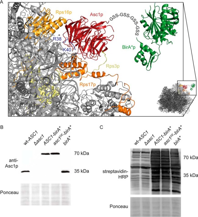
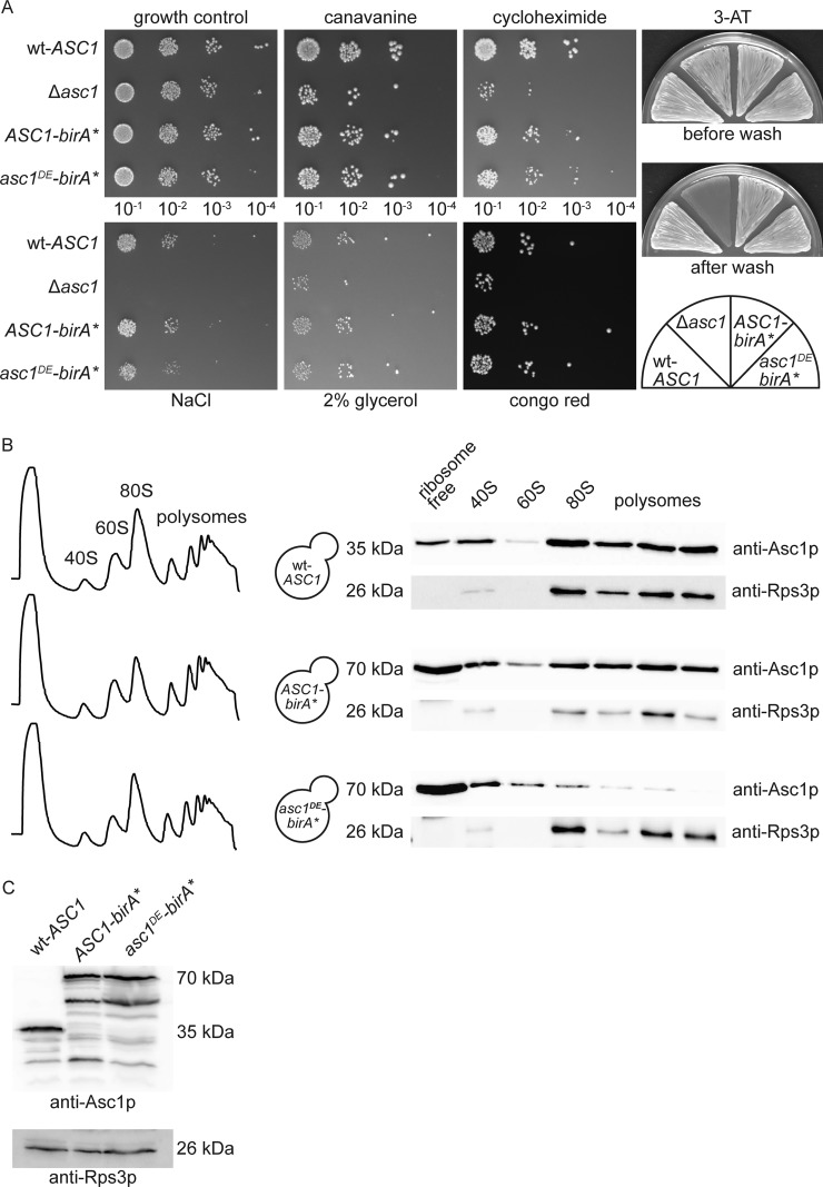
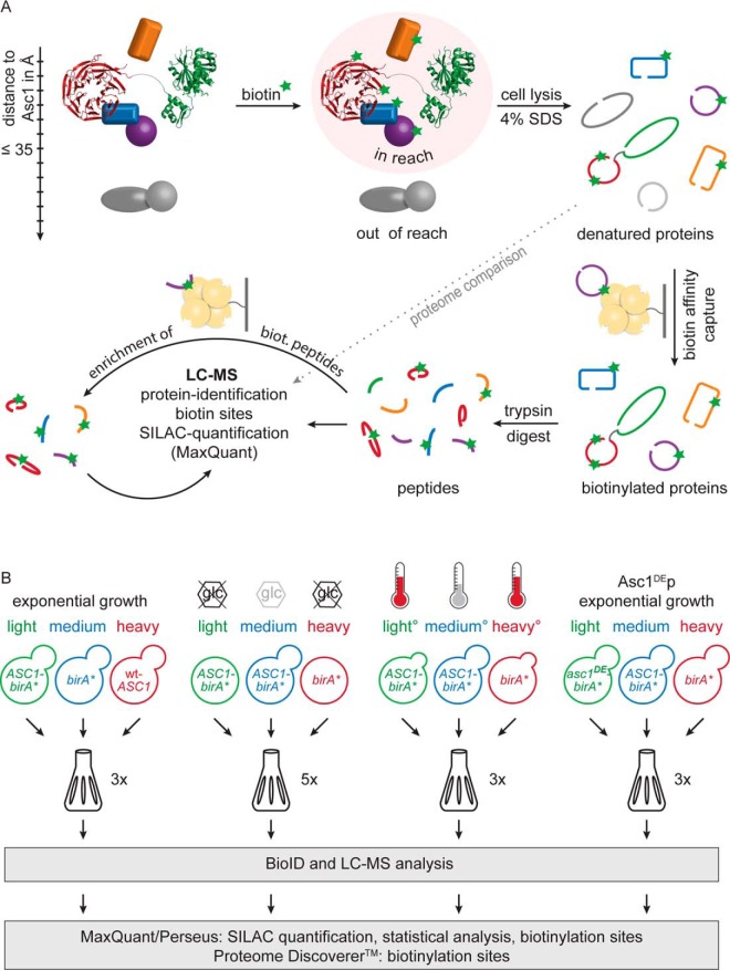
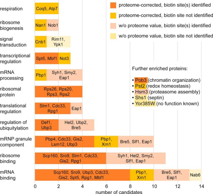
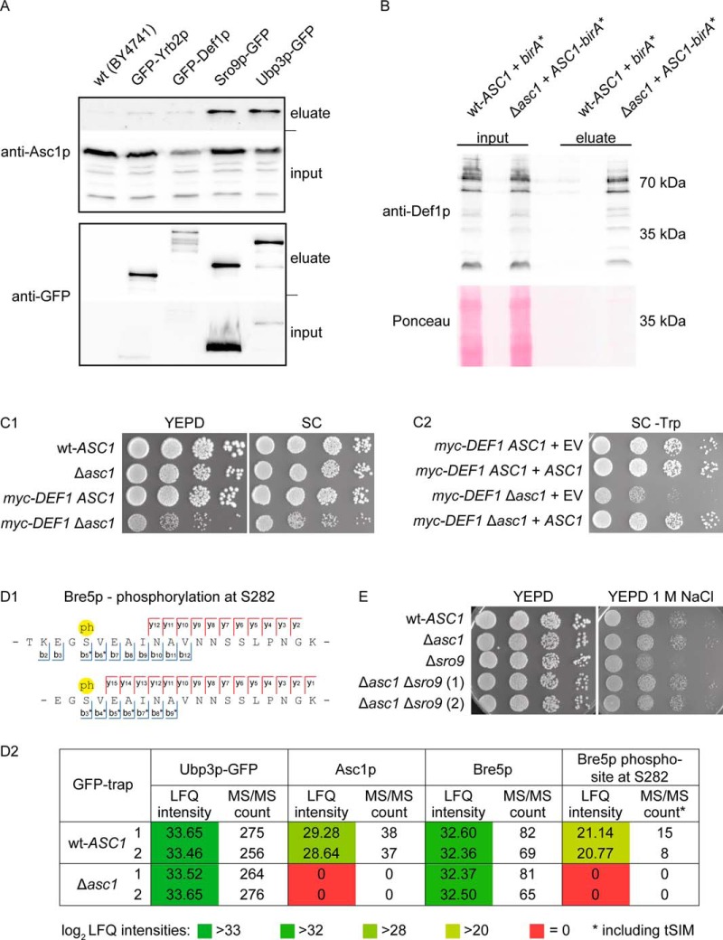
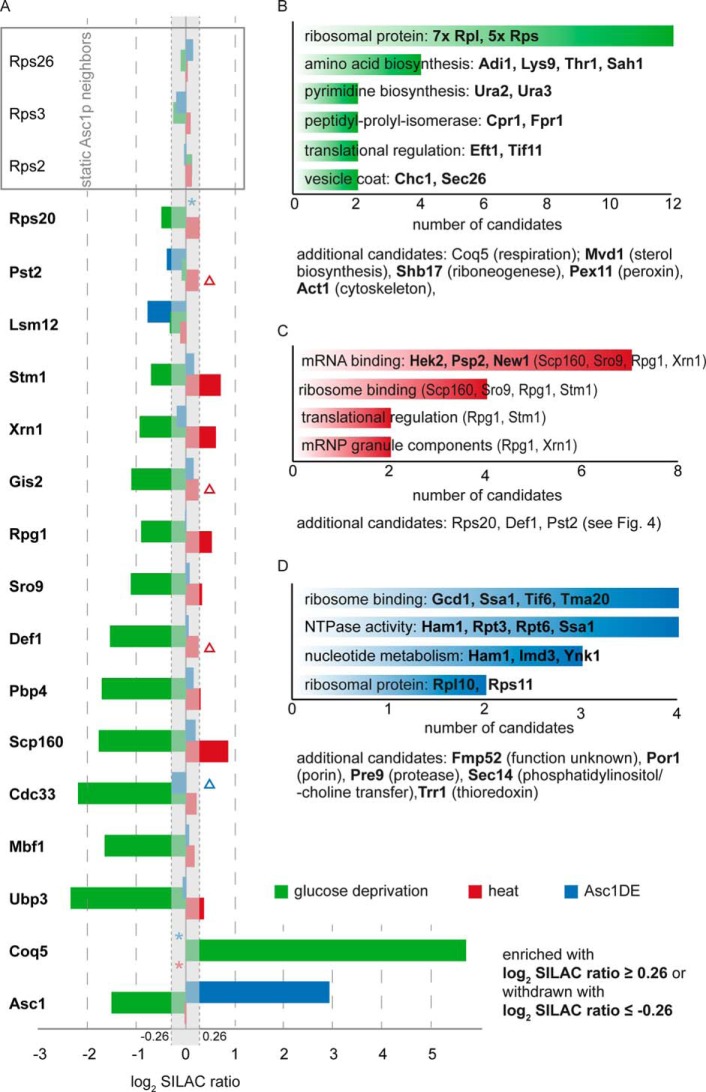
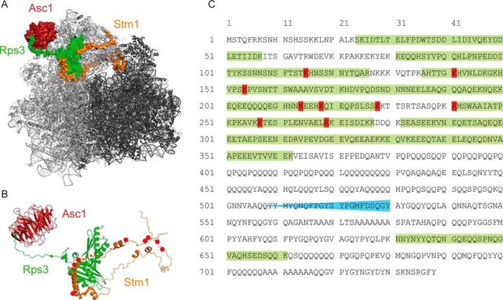
The Asc1 protein of Saccharomyces cerevisiae is a scaffold protein at the head region of ribosomal 40S that links mRNA translation to cellular signaling. In this study, proteins that colocalize with Asc1p were identified with proximity-dependent Biotin IDentification (BioID), an in vivo labeling technique described here for the first time for yeast. Biotinylated Asc1p-birA*-proximal proteins were identified and quantitatively verified against controls applying SILAC and mass spectrometry. The mRNA-binding proteins Sro9p and Gis2p appeared together with Scp160p, each providing ribosomes with nuclear transcripts. The cap-binding protein eIF4E (Cdc33p) and the eIF3/a-subunit (Rpg1p) were identified reflecting the encounter of proteins involved in the initiation of mRNA translation at the head region of ribosomal 40S. Unexpectedly, a protein involved in ribosome preservation (the clamping factor Stm1p), the deubiquitylation complex Ubp3p-Bre5p, the RNA polymerase II degradation factor 1 (Def1p), and transcription factors (Spt5p, Mbf1p) colocalize with Asc1p in exponentially growing cells. For Asc1R38D, K40Ep, a variant considered to be deficient in binding to ribosomes, BioID revealed its predominant ribosome localization. Glucose depletion replaced most of the Asc1p colocalizing proteins for additional ribosomal proteins, suggesting a ribosome aggregation process during early nutrient limitation, possibly concomitant with ribosomal subunit clamping. Overall, the characterization of the Asc1p microenvironment with BioID confirmed and substantiated our recent findings that the β-propeller broadly contributes to signal transduction influencing phosphorylation of colocalizing proteins (e.g. of Bre5p), and by that might affect nuclear gene transcription and the fate of ribosomes.


Figures

Fig. 1.
Fig. 1.
Scheme of Asc1-BirA*p at the head of the 40S ribosome. A, The Asc1 protein is a constituent of the 40S ribosomal subunit and interacts physically with the ribosomal proteins Rps3p, Rps16p and Rps17p. Amino acid residues Arg38 and Lys40 contribute to ribosome-binding and their exchange to Asp or Glu (Asc1DEp) weakens ribosome-binding. The BirA* protein is fused to the C-terminus of Asc1p and Asc1DEp via four repeats of a Gly-Ser-Ser linker sequence indicated as letter sequence. Because of the ribosome averted orientation of the Asc1p C-terminus, ribosome binding of the fusion proteins should not be sterically compromised. The crystal structure data of the S. cerevisiae 80S ribosome and the E. coli BirA protein derive from the PDB entries 4V88 (10) and 1BIB (73) and were combined to model the fusion protein with the PyMOL Molecular Graphics System software. B, Expression of the Asc1-BirA* and the Asc1DE-BirA* fusion proteins (∼70 kDa) from a high copy number plasmid in the Δasc1 strain background provides wild-type-like Asc1p levels. Proteins were detected in Western experiments using an Asc1p-specific antibody. The Ponceau staining of the lane is shown for a small part of the lanes. C, Protein biotinylation, detected with horseradish peroxidase (HRP)-coupled streptavidin, was elevated in strains expressing Asc1-BirA*p, Asc1DE-BirA*p or the mere BirA* protein cultivated in the presence of biotin. Parts of the Ponceau stained lanes are shown.
Fig. 2.
Fig. 2.
ASC1-birA* and asc1DE-birA* complement asc1 phenotypes and the fusion proteins associate with translating ribosomes. A, For drop dilution assays 10-fold dilution series of wt-ASC1, Δasc1, ASC1-birA* and asc1DE-birA* cell suspensions were spotted on YNB agar plates with the protein misfolding agent canavanine (600 ng/ml) or the translation inhibitor cycloheximide (0.15 μg/ml), or with NaCl (75 mm). Additionally, cells were dropped on YNB plates with 2% glycerol instead of glucose and on medium with congo red (125 μg/ml). A YNB agar plate was used for the growth control, and all plates were photographed after 3 to 4 days of growth. Adhesive growth was induced with 1 mm 3-AT and scored after 3 days of growth followed by gentle washing. ASC1-birA* and asc1DE-birA* were expressed from high copy number plasmids in the Δasc1 strain background. The wt-ASC1 and Δasc1 strains were transformed with an empty vector (pME2787). B, The ribosome-binding ability of the Asc1-BirA* and Asc1DE-BirA* fusion proteins in comparison to Asc1p was analyzed with sucrose gradient centrifugation and Western blot analysis with an Asc1p-specific antibody. The abundances of the different Asc1p variants in the nonribosomal fraction, the 40S and 60S subunits as well as the 80S monosome and the polysomes revealed ribosome-binding of the Asc1-BirA* protein, but ribosome dissociation of Asc1DE-BirA*p during ultracentrifugation. The distribution of Rps3p within these fractions was visualized with an Rps3p-specific antibody as marker for the migration of the small ribosomal subunit. C, Input loading control verified by Western blotting and detection with the Asc1p- and Rps3p-specific antibodies.
Fig. 3.
Fig. 3.
SILAC-BioID: Enrichment quantified against controls. A, Supplemented biotin was used by the Asc1-BirA* fusion protein to covalently biotinylate proteins in reach (up to ∼35 Å). After cell lysis in the presence of 4% SDS biotinylated proteins were enriched via biotin affinity capture and afterwards digested with trypsin. The resulting peptides were subjected to LC-MS analysis for protein identification and for the determination of biotinylated lysine residues. SILAC quantification was performed with the MaxQuant software (39). Additional database search with the Proteome Discoverer software enhanced the identification of biotinylated sites. To improve the coverage of biotinylated peptides, half of the peptide solution was subjected to additional biotin affinity capture and LC-MS analysis. Peptide samples without biotin affinity capture were also analyzed by LC-MS. The resulting proteome values were used to normalize affinity capture SILAC ratios against total protein abundances. Modified from Smolinski and Valerius (74). B, All four BioID-experiments were performed with triple SILAC to quantitatively compare cells of three different cultures in one batch. The birA* and wt-ASC1 strains served as negative controls. Alterations in the Asc1p-neighborhood on glucose starvation and heat stress (37 °C) were determined against ASC1-birA* at exponential growth (2% glucose, 30 °C) as reference and birA* at stress as negative control. The neighborhood of Asc1DE-BirA*p was compared with ASC1-birA* as reference and birA* as negative control. Yeast strains were individually cultivated in the presence of 10 μm biotin and were labeled with lysine and arginine isotope variants as indicated. All genes were expressed from high copy number plasmids in a Δarg4 Δlys1 strain background to ensure exclusive incorporation of the labeled amino acids into all proteins. The number of biological replicates is indicated for each experiment. °One replicate of the “heat stress” BioID experiment was performed with a label swap: ASC1-birA*, 37 °C (heavy), ASC1-birA*, 30 °C (medium), birA*, 37 °C (light).
Fig. 4.
Fig. 4.
Functional grouping of Asc1p-neighbors. 40 proteins identified in the Asc1p-vicinity were assigned to groups reflecting molecular functions in a nonexclusive manner. The number of proteins per group is given by the x axis. The number of potential Asc1p-neighbors per group with or without proteome value, and with or without identified biotin site is reflected by the respective colors. Corresponding candidates are listed.
Fig. 5.
Fig. 5.
Validation of selected BioID candidates. A, GFP-traps. GFP-tagged Def1p, Sro9p, Ubp3p, and Yrb2p (as a negative control) were enriched from the respective strains expressing the fusion-proteins. The wt strain BY4741 expresses no GFP and was used for a second negative control. Input and eluate fractions were subjected to Western blot experiments for the subsequent detection of Asc1p and GFP-fusion proteins with the respective antibodies. B, BioID Western. birA* and ASC1-birA* were expressed from high copy number plasmids in the wt-ASC1 and Δasc1 strains, respectively. The strains were cultivated in the presence of biotin, and biotinylated proteins were enriched from cell lysates of these cultures. The abundance of Def1p in the input control samples and the eluate fractions was analyzed with Western blot experiments using a Def1p-specific antibody. C, Genetic interaction of ASC1 and DEF1. (C1) Synthetic growth defect. Ten-fold dilution series of W303 wt-ASC1, Δasc1, myc-DEF1 ASC1, and myc-DEF1 Δasc1 cell suspensions were spotted on YEPD and SC medium. (C2) Complementation of the synthetic growth defect. myc-DEF1 ASC1 and myc-DEF1 Δasc1 cells were transformed with centromere plasmids either expressing ASC1 from its own promoter (pME4481) or without any ASC1 gene (EV, pME2781) as control. Cell suspensions were spotted onto SC medium without tryptophan (-Trp) for selection. D, Asc1p-dependent phosphorylation of Ubp3p-associated Bre5p. (D1) Bre5p phospho-peptides identified by mass spectrometry. Bre5p phospho-peptides with phosphorylation (ph) at S282 identified from tryptic digests of proteins enriched with Ubp3p-GFP. Both peptides cover the amino acid sequence 280 to 298. Because of a missed cleavage site C-terminal of K279, the upper peptide starts at T278. See supplemental Figs. S2 and S3 for the corresponding fragmentation spectra. The b- and y-ions found in these spectra are indicated. Highest phospho-site probabilities for S282: 100% (short peptide) and 93% (long peptide). (D2) Label-free quantification (LFQ) of proteins/peptides copurified with Ubp3p-GFP in the absence and presence of Asc1p. The experiment was performed with two biological replicates (1 and 2). LFQ intensities and MS/MS counts for Ubp3p-GFP, Asc1p, Bre5p, and the Bre5p phospho-site S282 are depicted. MS/MS counts for Bre5p phosphorylation at S282 derived from peptides depicted in D1 including tSIM analyses. E, Genetic interaction of ASC1 and SRO9. 10-fold dilution series of BY wt-ASC1, Δasc1, Δsro9, and Δasc1 Δsro9 cell suspensions were spotted on YEPD plates with and without 1 m NaCl. Two Δasc1 Δsro9 clones were tested. Strains of the S288c background are less osmosensitive then strains of the Σ1278b background; therefore, an increased osmosensitivity of the Δasc1 strain was not observed here (compare with Fig. 2).
Fig. 6.
Fig. 6.
Changes within the proteinaceous neighborhood of Asc1p. A, The bar chart depicts changes in the Asc1-BirA*p neighborhood in cells during glucose deprivation and heat stress (37 °C), and because of the R38D K40E exchanges relative to the neighborhood of Asc1-BirA*p during exponential growth. 19 proteins identified as Asc1p-neighbors during exponential growth are listed on the left. The bars represent log2 SILAC ratios/enrichment values of the ASC1-birA*-strain starved from glucose (green), cultivated at 37 °C (red) or of the ASC1DE-birA*-strain (blue). Proteins were considered as dynamic Asc1p-neighbors if log2 SILAC ratios were outside −0.26 and 0.26. Stable Asc1p-neighbors, independent of the tested growth conditions or the DE-exchange, are listed at the top within the gray box (log2 SILAC ratio between −0.26 and 0.26). Missing SILAC ratios are indicated by asterisks. Mean values that do not reach the respective threshold because of outlier values are indicated by triangles. B–D, Proteins specifically enriched in the Asc1p-neighborhood at (B) glucose starvation, (C) heat-stress, or (D) of the Asc1DEp-variant were assigned to functional groups in a nonexclusive manner. Candidates of the respective groups are listed within the bar chart, those that are exclusively enriched in these specific experiments are listed in bold. The number of proteins per group is given by the x axis. Further proteins that do not belong to the chosen groups are listed below the diagrams.
Fig. 7.
Fig. 7.
Stm1p and Def1p are biotinylated by the Asc1-BirA* fusion protein at several sites. A, At glucose deprivation the ribosomal clamping factor Stm1p is located in between the 40S and 60S subunits and prevents mRNA translation. At this condition, Asc1p forms a tightly connected network with Rps3p and Stm1p at the ribosome. B, Stm1p appears as a protein with almost no intrinsic structure during glucose starvation. Biotinylated lysine residues are indicated by red dots. The crystal structure data derive from the PDB entry 4V88 (10) and were used for visualization with the PyMOL Molecular Graphics System software. C, Def1p was biotinylated mainly in the N-terminal half of the protein. Proteasome-dependent processing of Def1p in response to transcription stress occurs before amino acid 530, and the presence of a C-terminal peptide confirms cytoplasmic proximity of Def1p to Asc1p. Biotinylated lysine residues are colored in red. The area of Def1p-processing is indicated in blue and peptides identified by LC-MS analysis are highlighted in green.

References

    1. Pan C. Q., Sudol M., Sheetz M., and Low B. C. (2012) Modularity and functional plasticity of scaffold proteins as p(l) acemakers in cell signaling. Cell Signal 24, 2143–2165 - PubMed
    1. Vondriska T. M., Pass J. M., and Ping P. (2004) Scaffold proteins and assembly of multiprotein signaling complexes. J. Mol. Cell Cardiol 37, 391–397 - PubMed
    1. Chantrel Y., Gaisne M., Lions C., and Verdière J. (1998) The transcriptional regulator hap1p (cyp1p) is essential for anaerobic or heme-deficient growth of saccharomyces cerevisiae: genetic and molecular characterization of an extragenic suppressor that encodes a wd repeat protein. Genetics 148, 559–569 - PMC - PubMed
    1. Coyle S. M., Gilbert W. V., and Doudna J. A. (2009) Direct link between rack1 function and localization at the ribosome in vivo. Mol. Cell. Biol. 29, 1626–1634 - PMC - PubMed
    1. Tarnowski K., Fituch K., Szczepanowski R. H., Dadlez M., and Kaus-Drobek M. (2014) Patterns of structural dynamics in rack1 protein retained throughout evolution: a hydrogen-deuterium exchange study of three orthologs. Protein Sci. Publ. Protein. Soc. 23, 639–651 - PMC - PubMed

MeSH terms

Substances

LinkOut - more resources