Review
. 2018 Feb;39(2):208-216.
doi: 10.3174/ajnr.A5391. Epub 2017 Oct 5.

Radiomics in Brain Tumor: Image Assessment, Quantitative Feature Descriptors, and Machine-Learning Approaches

Affiliations
Review

Radiomics in Brain Tumor: Image Assessment, Quantitative Feature Descriptors, and Machine-Learning Approaches

M Zhou et al. AJNR Am J Neuroradiol. 2018 Feb.

Abstract

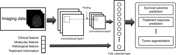
Radiomics describes a broad set of computational methods that extract quantitative features from radiographic images. The resulting features can be used to inform imaging diagnosis, prognosis, and therapy response in oncology. However, major challenges remain for methodologic developments to optimize feature extraction and provide rapid information flow in clinical settings. Equally important, to be clinically useful, predictive radiomic properties must be clearly linked to meaningful biologic characteristics and qualitative imaging properties familiar to radiologists. Here we use a cross-disciplinary approach to highlight studies in radiomics. We review brain tumor radiologic studies (eg, imaging interpretation) through computational models (eg, computer vision and machine learning) that provide novel clinical insights. We outline current quantitative image feature extraction and prediction strategies with different levels of available clinical classes for supporting clinical decision-making. We further discuss machine-learning challenges and data opportunities to advance radiomic studies.


Figures

Fig 1.
Fig 1.
Visualization of computational image feature descriptors. A T1-weighted brain tumor section (A and B) is displayed, and feature visualizations (C–E) are given of LBP (C), HOG (D), and SIFT (E) descriptors. LBP quantifies local pixel structures through a binary coding scheme. HOG computes block-wise histogram gradients with multiple orientations. SIFT detects distributed key points with radius on tumor images. These multiparametric features create a rich image-driven data base to characterize tumors in MR imaging at different scales.
Fig 2.
Fig 2.
Linking subregional imaging to molecular profiles in glioblastoma. In this example, tumor subregions (B) are defined by jointly clustering on contrast-enhanced T1WI and T2WI (A). These subregions correspond to red (high T1WI and high T2WI), yellow (high T1WI and low T2WI), blue (low T1WI and high T2WI), and pink (low T1WI and low T2WI) areas. The defined tumor subregions enable quantitative spatial characterization, offering a means to noninvasively assess specific molecular activities (C) with enriched molecular pathways (D).
Fig 3.
Fig 3.
Illustration of the convolutional neural networks model using imaging and other biomedical data for brain tumor analysis. The convolutional neural networks model consists of multiple convolutional layers, pooling layers, and fully connected layers to learn an abstraction of the input data, such as imaging and clinical features for a variety of outcome evaluations.

References

    1. Gillies RJ, Kinahan PE, Hricak H. Radiomics: images are more than pictures, they are data. Radiology 2016;278:563–77 10.1148/radiol.2015151169 - DOI - PMC - PubMed
    1. Kumar V, Gu YH, Basu S, et al. Radiomics: the process and the challenges. Magn Reson Imaging 2012;30:1234–48 10.1016/j.mri.2012.06.010 - DOI - PMC - PubMed
    1. Ludwig JA, Weinstein JN. Biomarkers in cancer staging, prognosis and treatment selection. Nat Rev Cancer 2005;5:845–56 10.1038/nrc1739 - DOI - PubMed
    1. Prior FW, Clark K, Commean P, et al. TCIA: an information resource to enable open science. In: Proceedings of the 35th Annual International Conference of the IEEE Engineering in Medicine and Biology Society, Osaka, Japan July 3–7, 2013:1282–85 - PMC - PubMed
    1. Zhou M, Chaudhury B, Hall LO, et al. Identifying spatial imaging biomarkers of glioblastoma multiforme for survival group prediction. J Magn Reson Imaging 2016;46:115–23 - PubMed

Publication types

MeSH terms

LinkOut - more resources