. 2017 Oct 5;7(1):12707.
doi: 10.1038/s41598-017-11880-z.

Therapeutic potential of human minor salivary gland epithelial progenitor cells in liver regeneration

Affiliations

Therapeutic potential of human minor salivary gland epithelial progenitor cells in liver regeneration

Chen Zhang et al. Sci Rep. .

Abstract

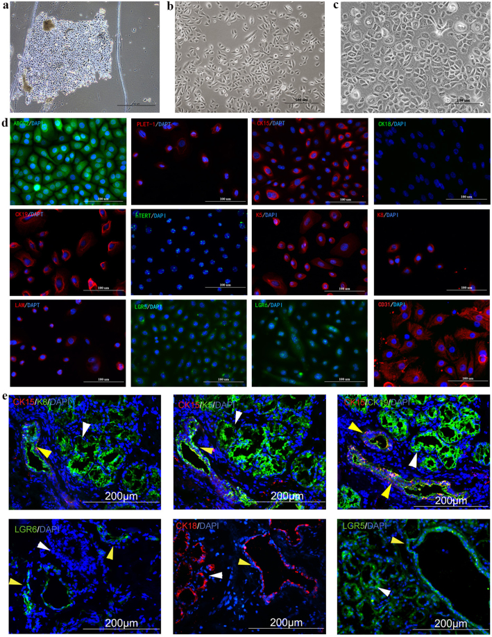
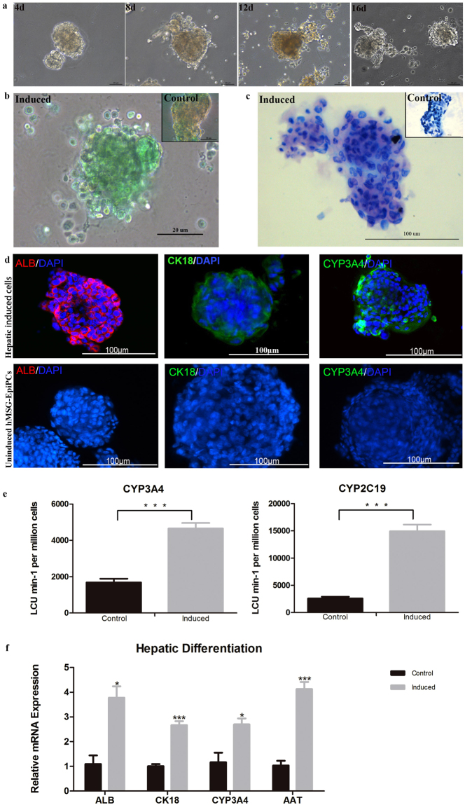
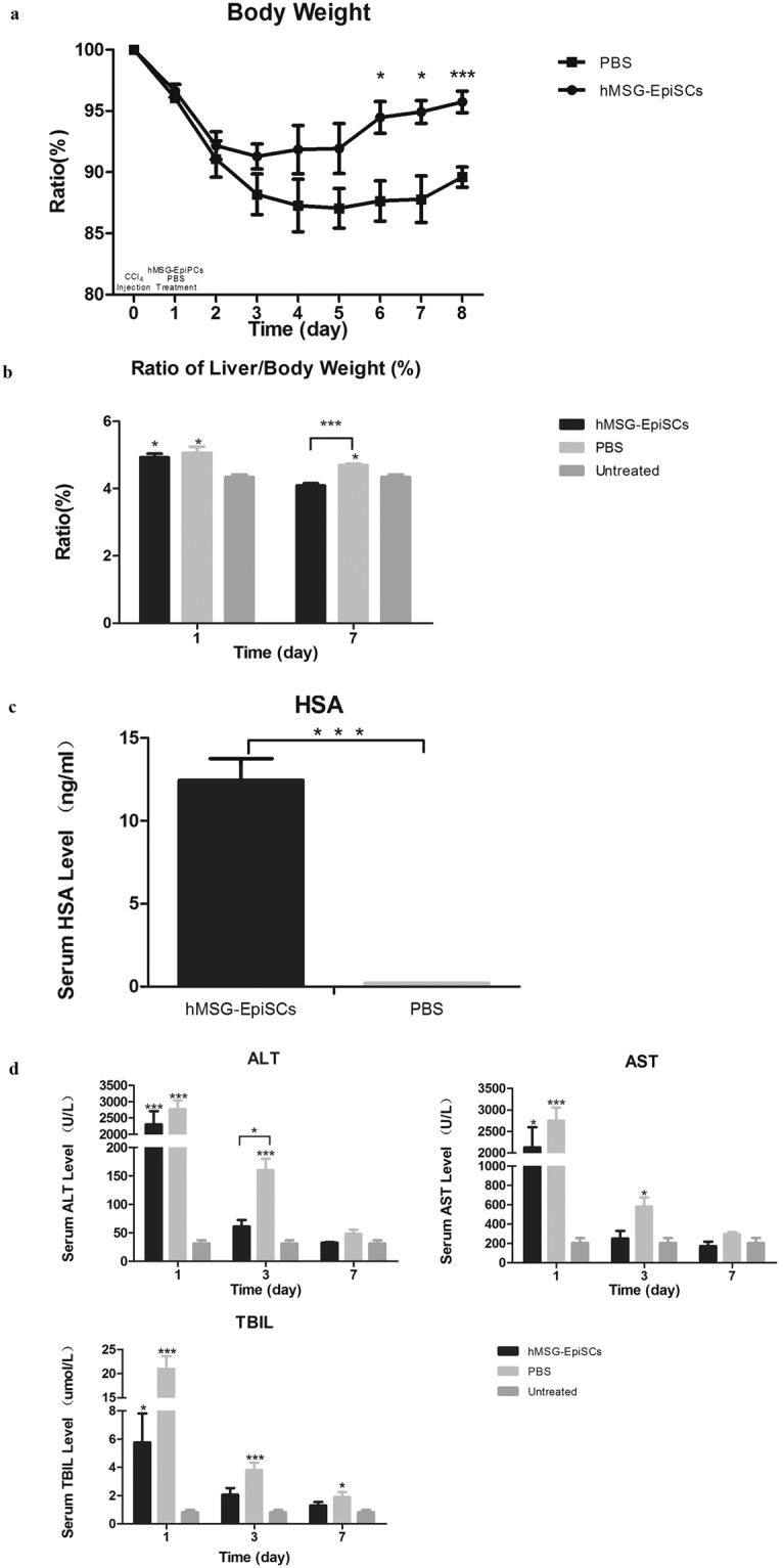
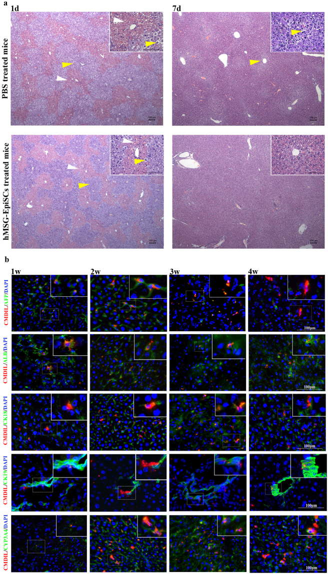
Liver disease is a serious problem affecting millions of people with continually increasing prevalence. Stem cell therapy has become a promising treatment for liver dysfunction. We previously reported on human minor salivary gland mesenchymal stem cells (hMSGMSCs), which are highly self-renewable with multi-potent differentiation capability. In this study, keratinocyte-like cells with self-regeneration and hepatic differentiation potential were isolated and characterized, and named human minor salivary gland epithelial progenitor cells (hMSG-EpiPCs). hMSG-EpiPCs were easily obtained via minor intraoral incision; they expressed epithelial progenitor/stem cell and other tissue stem cell markers such as CD29, CD49f, cytokeratins, ABCG2, PLET-1, salivary epithelial cell markers CD44 and CD166, and the Wnt target related gene LGR5 and LGR6. The cells were induced into functional hepatocytes in vitro which expressed liver-associated markers ALB, CYP3A4, AAT, and CK18. Upon transplantation in vivo, they ameliorated severe acute liver damage in SCID mice caused by carbon tetrachloride (CCl4) injection. In a two-thirds partial hepatectomy mouse model, the transplanted cells survived at least 4 weeks and exhibited hepatic potential. These findings demonstrate that hMSG-EpiPCs have potential as a cellular therapy basis for hepatic diseases, physiological and toxicology studies and regenerative medicine.


Conflict of interest statement

The authors declare that they have no competing interests.

Figures

Figure 1
Figure 1
Culture and Characterization of hMSG-EpiPCs. (a) Primary culture for hMSG-EpiPCs at day 5. (b) Expanded hMSG-EpiPCs of passage 3. (c) Expanded hMSG-EpiPCs of passage 25. (d) Immunostaining of expanded hMSG-EpiPCs of passage 5. (e) Immunostaining of hMSG tissues (Yellow arrows: excretory duct, white arrows: acinus).
Figure 2
Figure 2
Hepatic differentiation of hMSG-EpiPCs in vitro. (a) Picture of induced hMSG-EpiPCs taken on day 4, 8, 12 and 16 from left to right. (b) Indocyanine green (ICG) uptake assay on day 16 of hepatic induction and the control group. (c) Periodic acid-Schiff (PAS) staining on day 16 of hepatic induction and the control group. (d) Immunostaining on day 16 of hepatic induced and uninduced cells for ALB, CK18 and CYP3A4 from left to right. (e) CYP450 activity analysis on day 16 of hepatic induction for CYP3A4 and CYP2C19. (f) Quantitative real-time PCR analysis of relative mRNA expression of hMSG-EpiPCs and induced hepatic cells on day 16. (***P < 0.01; n ≧ 3).
Figure 3
Figure 3
hMSG-EpiPCs treatment promotes liver regeneration after acute injury. (a) Changes in body weight of CCl4-injected mice show that hMSG-EpiPCs treatment group restored body weight faster than control group. (b) The ratio of liver weight to body weight of the hMSG-EpiPCs treatment mice was lower than that of the PBS-treated mice. (c) HSA could be detected in hMSG-EpiPCs treatment group. (d) Serum ALT,AST and TBIL level increased in all mice 2 day after CCl4 injection, and decreased faster in hMSG-EpiPCs treatment group than that of control mice. (*P < 0.05; ***P < 0.01, n ≧ 3).
Figure 4
Figure 4
Hepatic differentiation of hMSG-EpiPCs in vivo. (a) H&E staining of mice liver section on day 1 and 7 post PBS and hMSG-EpiPCs injection (White arrows: erythrocyte diapedesis, yellow arrows: diseased hepatocytes). (b) Immunostaining 1, 2, 3 and 4 weeks after hMSG-EpiPCs transplantation from left to right, green fluorescence signal for AFP, ALB, CK18, CK19 and CYP3A4 from top to bottom, and red for CM-Dil labeling.

References

    1. Pai M, et al. Autologous infusion of expanded mobilized adult bone marrow-derived CD34+ cells into patients with alcoholic liver cirrhosis. The American journal of gastroenterology. 2008;103:1952–1958. doi: 10.1111/j.1572-0241.2008.01993.x. - DOI - PubMed
    1. Terai S, et al. Improved liver function in patients with liver cirrhosis after autologous bone marrow cell infusion therapy. Stem cells. 2006;24:2292–2298. doi: 10.1634/stemcells.2005-0542. - DOI - PubMed
    1. Vassilopoulos G, Wang PR, Russell DW. Transplanted bone marrow regenerates liver by cell fusion. Nature. 2003;422:901–904. doi: 10.1038/nature01539. - DOI - PubMed
    1. Peng L, et al. Autologous bone marrow mesenchymal stem cell transplantation in liver failure patients caused by hepatitis B: Short-term and long-term outcomes. Hepatology. 2011;54:820–828. doi: 10.1002/hep.24434. - DOI - PubMed
    1. Apel A, et al. Suitability of human mesenchymal stem cells for gene therapy depends on the expansion medium. Experimental Cell Research. 2009. 2009;315:498–507. doi: 10.1016/j.yexcr.2008.11.013. - DOI - PubMed

MeSH terms

Substances