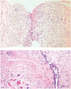
Desmoid Tumors: A Clear Perspective or a Persisting Enigma? A Case Report and Review of Literature
- PMID: 28983359
- PMCID: PMC5624685
- DOI: 10.14740/wjon961w
Desmoid Tumors: A Clear Perspective or a Persisting Enigma? A Case Report and Review of Literature
Abstract
Desmoid tumors are benign but locally aggressive tumors of mesenchymal origin which are poorly circumscribed, infiltrate the surrounding tissue, lack a true capsule and are composed of abundant collagen. History of trauma or surgery to the site of tumor origin is elicited in up to one in four cases and they most commonly develop in the anterior abdominal wall and shoulder girdle but they can arise in any skeletal muscle. The clinical behavior and natural history of desmoid tumors are unpredictable and management is difficult with many issues remaining controversial, mainly regarding early detection, the role, type and timing of surgery and the value of non-operative therapies. We report a case of anterior abdominal wall desmoid tumor in a 40-year-old male with a previous history of surgery.
Keywords: Adenomatous polyposis coli mutation; Desmoid; Familial adenomatous polyposis; Gardner’s syndrome; Mesenchymal tumors.
Conflict of interest statement
None.
Figures


Similar articles
-
A massive abdominal wall desmoid tumor occurring in a laparotomy scar: a case report.World J Surg Oncol. 2011 Mar 22;9:35. doi: 10.1186/1477-7819-9-35. World J Surg Oncol. 2011. PMID: 21426541 Free PMC article.
-
Reconstruction of an abdominal wall defect with biologic mesh after resection of a desmoid tumor in a patient with a Gardner's syndrome.Acta Chir Belg. 2017 Feb;117(1):55-60. doi: 10.1080/00015458.2016.1212499. Epub 2016 Aug 18. Acta Chir Belg. 2017. PMID: 27538186
-
Surgery, desmoid tumors, and familial adenomatous polyposis: case report and literature review.Am J Gastroenterol. 1996 Dec;91(12):2598-601. Am J Gastroenterol. 1996. PMID: 8946994 Review.
-
Family history, surgery, and APC mutation are risk factors for desmoid tumors in familial adenomatous polyposis: an international cohort study.Dis Colon Rectum. 2011 Oct;54(10):1229-34. doi: 10.1097/DCR.0b013e318227e4e8. Dis Colon Rectum. 2011. PMID: 21904137
-
[Long-term experience with therapy of a female patient with Gardner's syndrome, first presenting with extra-abdominal desmoid tumor, and review of the literature].Magy Seb. 2009 Apr;62(2):75-82. doi: 10.1556/MaSeb.62.2009.2.5. Magy Seb. 2009. PMID: 19386568 Review. Hungarian.
Cited by
-
Pregnancy-Associated Giant Abdominal Desmoid Tumor: A Case Report of Active Surveillance and Surgical Management.Int J Womens Health. 2025 Jul 21;17:2227-2232. doi: 10.2147/IJWH.S532226. eCollection 2025. Int J Womens Health. 2025. PMID: 40718085 Free PMC article.
-
Huge mesenteric desmoid-type fibromatosis with unusual presentation: A case report.Ann Med Surg (Lond). 2022 May 10;78:103741. doi: 10.1016/j.amsu.2022.103741. eCollection 2022 Jun. Ann Med Surg (Lond). 2022. PMID: 35600202 Free PMC article.
-
Retroperitoneal invasive fibromatosis after laparoscopic radical resection of colon cancer: a case report and literature review.Front Oncol. 2025 Jul 17;15:1582253. doi: 10.3389/fonc.2025.1582253. eCollection 2025. Front Oncol. 2025. PMID: 40746605 Free PMC article.
-
Retroperitoneal desmoid-type fibromatosis: a case report.Ann Med Surg (Lond). 2023 Apr 6;85(4):1258-1261. doi: 10.1097/MS9.0000000000000491. eCollection 2023 Apr. Ann Med Surg (Lond). 2023. PMID: 37113969 Free PMC article.
References
-
- Moslein G, Dozois RR. Desmoid tumors associated with familial adenomatous polyposis. Perspectives in Colon and Rectal Surgery. 1998;10:109–126.
-
- Sturt NJ, Clark SK. Current ideas in desmoid tumours. Fam Cancer. 2006;5(3):275–285. discussion 287–278. - PubMed
Publication types
LinkOut - more resources
Full Text Sources
Other Literature Sources
Miscellaneous
