. 2017 Jul;4(3):035002.
doi: 10.1117/1.NPh.4.3.035002. Epub 2017 Sep 27.

Characterization of a fiber-less, multichannel optical probe for continuous wave functional near-infrared spectroscopy based on silicon photomultipliers detectors: in-vivo assessment of primary sensorimotor response

Affiliations

Characterization of a fiber-less, multichannel optical probe for continuous wave functional near-infrared spectroscopy based on silicon photomultipliers detectors: in-vivo assessment of primary sensorimotor response

Antonio M Chiarelli et al. Neurophotonics. 2017 Jul.

Abstract

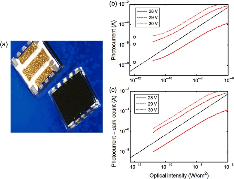
We report development, testing, and in vivo characterization of a multichannel optical probe for continuous wave (CW) functional near-infrared spectroscopy (fNIRS) that relies on silicon photomultipliers (SiPMs) detectors. SiPMs are cheap, low voltage, and robust semiconductor light detectors with performances analogous to photomultiplier tubes (PMTs). In contrast with PMTs, SiPMs allow direct contact with the head and transfer of the analog signals through thin cables greatly increasing the system flexibility avoiding optical fibers. The coupling of SiPMs and light-emitting diodes (LEDs) made the optical probe lightweight and robust against motion artifacts. After characterization of SiPM performances, which was proven to provide a noise equivalent power below 3 fW, the apparatus was compared through an in vivo experiment to a commercial system relying on laser diodes, PMTs, and optical fibers for light probing and detection. The optical probes were located over the primary sensorimotor cortex and the similarities between the hemodynamic responses to the contralateral motor task were assessed. When compared to other state-of-the-art wearable fNIRS systems, where photodiode detectors are employed, the single photon sensitivity and dynamic range of SiPMs can fully exploit the long and variable interoptode distances needed for correct estimation of brain hemodynamics using CW-fNIRS.

Keywords: continuous wave functional near-infrared spectroscopy; primary sensorimotor response; silicon photomultipliers; wearable neuroimaging.


Figures

Fig. 1
Fig. 1
(a) Example of two large area N-on-P SiPM detectors manufactured at ST (front and back view). The devices are packaged in a surface mount device (SMD) housing with 5.1×5.1  mm2 total area. (b) SiPMs photocurrent as a function of optical intensity measured at 25°C. Dark count levels and photocurrents at three bias voltages are reported as open circles, and as continuous, dashed, and dotted red lines, respectively. The ideal linear trend is indicated by the dashed black line. (c) SiPMs photocurrents after subtraction of the dark count rate. Photocurrents are reported as continuous, dashed, and dotted red lines. The ideal linear trend is indicated by the dashed black line.
Fig. 2
Fig. 2
(a) Example of a SIPM and a LED mounted on the small boards that were connected to the acquisition system and that were employed in the final dark-fabric optical probe. (b) Schematic representation of the optodes’ layout employed.
Fig. 3
Fig. 3
Electrical scheme of the system.
Fig. 4
Fig. 4
(a) Average SNR of the system at a fixed incident light power of 2 pW as a function of the sampling frequency of the system. (b) Estimated system’s NEP as a function of the sampling frequency. Data were collected in a range (1 to 20 Hz) compatible with actual fNIRS measurements.
Fig. 5
Fig. 5
Example of signals from 1 LED at 850 nm recorded by the three SIPMs of the optical probe employed. The data were taken in vivo and at rest, and they were estimated with an integration time of 10 ms. The average current was subtracted to expose physiological signals, such as heart rate and Mayer waves.
Fig. 6
Fig. 6
Average O2Hb and HHb responses to the motor task and related standard errors for the six channels and the two systems employed. The task period is identified in time by the two vertical black lines. Optodes locations on the scalp together with subject’s C3 location (10 to 20 system) are reported on a rendition of the subject’s magnetic resonance image. The blue arrows connect the midpoint of the optical channels on the rendered magnetic resonance image to their estimate of O2Hb and HHb responses.
Fig. 7
Fig. 7
(a) Schematization of the metric employed for system comparison. The metric of covariance between a standard hemodynamic response (left image) and the measured hemoglobin responses (right image) to the task was employed. (b) Scatterplot of covariances for the six channels and the two forms of hemoglobin (12 points) for one system (SiPM-LED) as a function of covariances of the commercial system (ISS Imagent™, Champaign, Illinois). (c) Bland and Altman plot of covariances of the two systems (SiPM-LED, ISS Imagent™). The differences in the covariance metric are reported as a function of the average of the covariance metric.

References

    1. Ferrari M., Quaresima V., “A brief review on the history of human functional near-infrared spectroscopy (fNIRS) development and fields of application,” Neuroimage 63(2), 921–935 (2012).NEIMEF10.1016/j.neuroimage.2012.03.049 - DOI - PubMed
    1. Villringer A., Chance B., “Non-invasive optical spectroscopy and imaging of human brain function,” Trends Neurosci. 20(10), 435–442 (1997).TNSCDR10.1016/S0166-2236(97)01132-6 - DOI - PubMed
    1. Giacometti P., Diamond S. G., “Correspondence of electroencephalography and near-infrared spectroscopy sensitivities to the cerebral cortex using a high-density layout,” Neurophotonics 1(2), 025001 (2014).10.1117/1.NPh.1.2.025001 - DOI - PMC - PubMed
    1. Raichle M. E., “Behind the scenes of functional brain imaging: a historical and physiological perspective,” Proc. Natl. Acad. Sci. U. S. A. 95(3), 765–772 (1998).10.1073/pnas.95.3.765 - DOI - PMC - PubMed
    1. Gratton G., Chiarelli A. M., Fabiani M., “From brain to blood vessels and back: a noninvasive optical imaging approach,” Neurophotonics 4(3), 031208 (2017).10.1117/1.NPh.4.3.031208 - DOI - PMC - PubMed