

Choriocapillaris Imaging Using Multiple En Face Optical Coherence Tomography Angiography Image Averaging
- PMID: 28983552
- PMCID: PMC5710392
- DOI: 10.1001/jamaophthalmol.2017.3904
Choriocapillaris Imaging Using Multiple En Face Optical Coherence Tomography Angiography Image Averaging
Abstract
Importance: Imaging of the choriocapillaris in vivo is challenging with existing technology. Optical coherence tomography angiography (OCTA), if optimized, could make the imaging less challenging.
Objective: To investigate multiple en face image averaging on OCTA images of the choriocapillaris.
Design, setting, and participants: Observational, cross-sectional case series at a referral institutional practice in Los Angeles, California. From the original cohort of 21 healthy individuals, 17 normal eyes of 17 participants were included in the study. The study dates were August to September 2016.
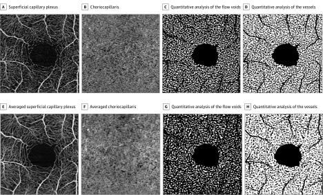
Exposures: All participants underwent OCTA imaging of the macula covering a 3 × 3-mm area using OCTA software (Cirrus 5000 with AngioPlex; Carl Zeiss Meditec). One eye per participant was repeatedly imaged to obtain 9 OCTA cube scan sets. Registration was first performed using superficial capillary plexus images, and this transformation was then applied to the choriocapillaris images. The 9 registered choriocapillaris images were then averaged. Quantitative parameters were measured on binarized OCTA images and compared with the unaveraged OCTA images.
Main outcome and measure: Vessel caliber measurement.
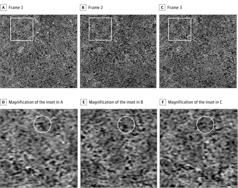
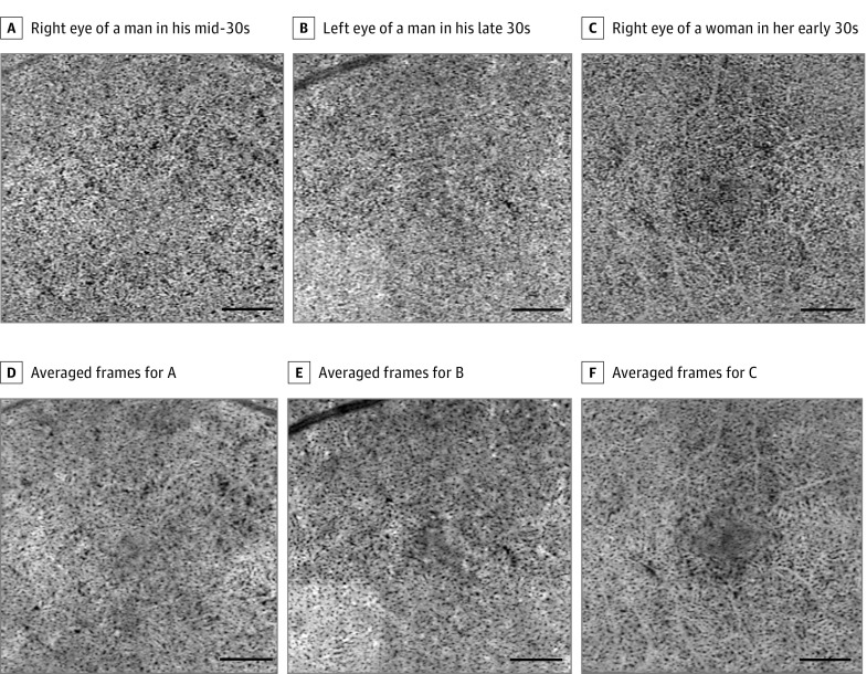
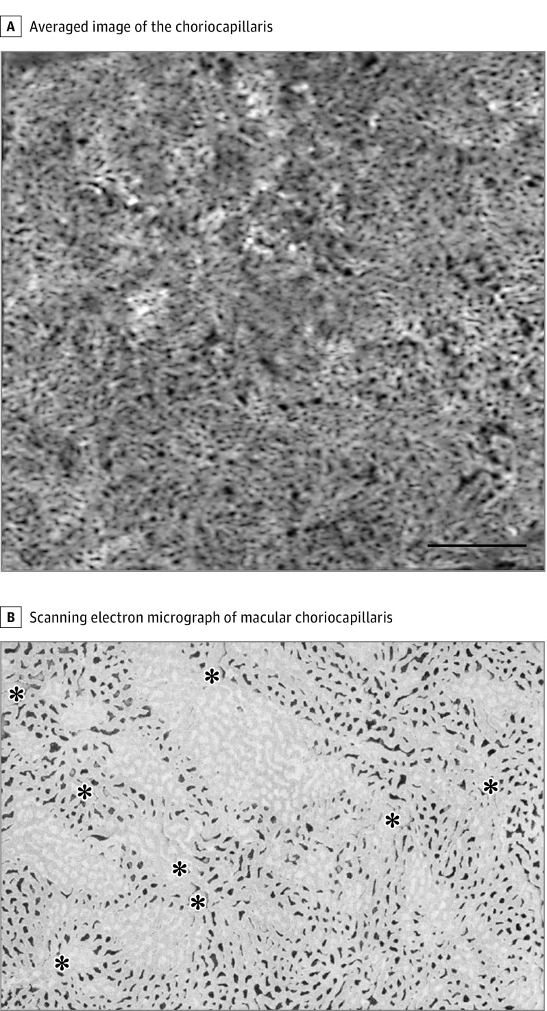
Results: Seventeen eyes of 17 participants (mean [SD] age, 35.1 [6.0] years; 9 [53%] female; and 9 [53%] of white race/ethnicity) with sufficient image quality were included in this analysis. The single unaveraged images demonstrated a granular appearance, and the vascular pattern was difficult to discern. After averaging, en face choriocapillaris images showed a meshwork appearance. The mean (SD) diameter of the vessels was 22.8 (5.8) µm (range, 9.6-40.2 µm). Compared with the single unaveraged images, the averaged images showed more flow voids (1423 flow voids [95% CI, 967-1909] vs 1254 flow voids [95% CI, 825-1683], P < .001), smaller average size of the flow voids (911 [95% CI, 301-1521] µm2 vs 1364 [95% CI, 645-2083] µm2, P < .001), and greater vessel density (70.7% [95% CI, 61.9%-79.5%] vs 61.9% [95% CI, 56.0%-67.8%], P < .001). The distribution of the number vs sizes of the flow voids was skewed in both unaveraged and averaged images. A linear log-log plot of the distribution showed a more homogeneous distribution in the averaged images compared with the unaveraged images.
Conclusions and relevance: Multiple en face averaging can improve visualization of the choriocapillaris on OCTA images, transforming the images from a granular appearance to a level where the intervascular spaces can be resolved in healthy volunteers.
Conflict of interest statement
Figures
Comment in
-
Image Averaging, a Powerful Tool in Optical Coherence Tomography and Optical Coherence Tomography Angiography.JAMA Ophthalmol. 2017 Nov 1;135(11):1204-1205. doi: 10.1001/jamaophthalmol.2017.4015. JAMA Ophthalmol. 2017. PMID: 28983576 No abstract available.
References
-
- Guyer DR, Schachat AP, Green WR The choroid: structural considerations. In: Ryan SJ, Schachat AP, Wilkinson CP, Hinton DR, eds. Retina. Vol 1. 4th ed Edinburgh, Scotland: Mosby; 2006:33-42.
-
- Olver JM. Functional anatomy of the choroidal circulation: methyl methacrylate casting of human choroid. Eye (Lond). 1990;4(pt 2):262-272. - PubMed
-
- Yoneya S, Tso MO. Angioarchitecture of the human choroid. Arch Ophthalmol. 1987;105(5):681-687. - PubMed
Publication types
MeSH terms
LinkOut - more resources
Full Text Sources
Other Literature Sources
Research Materials
