. 2017 Dec;16(6):8200-8208.
doi: 10.3892/mmr.2017.7648. Epub 2017 Sep 28.

Iguratimod prevents ovariectomy‑induced bone loss and suppresses osteoclastogenesis via inhibition of peroxisome proliferator‑activated receptor‑γ

Affiliations

Iguratimod prevents ovariectomy‑induced bone loss and suppresses osteoclastogenesis via inhibition of peroxisome proliferator‑activated receptor‑γ

Ying-Xing Wu et al. Mol Med Rep. 2017 Dec.

Abstract

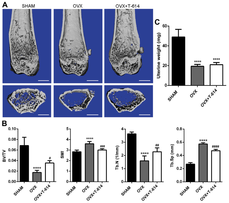
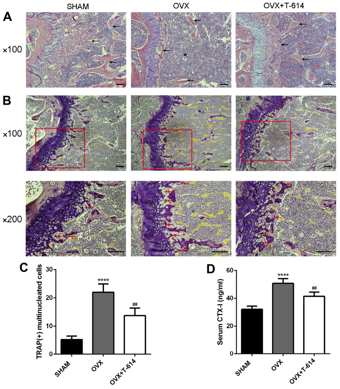
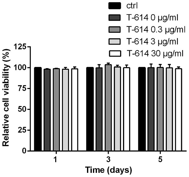
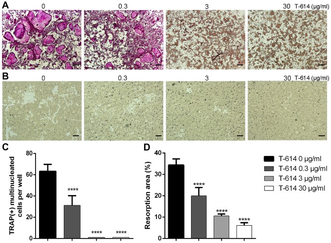
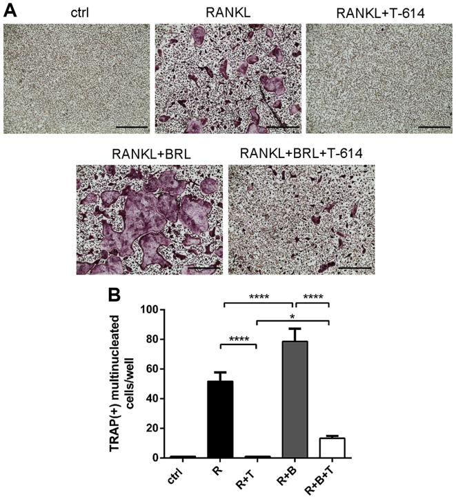
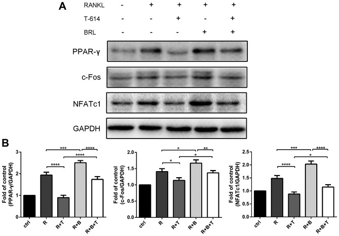
Iguratimod is known for its anti‑inflammatory activities and therapeutic effects in patients with rheumatoid arthritis. It has previously been demonstrated that iguratimod attenuates bone destruction and osteoclast formation in the Walker 256 rat mammary gland carcinoma cell‑induced bone cancer pain model. Therefore, it was hypothesized that iguratimod may additionally exhibit therapeutic effects on benign osteoclast‑associated diseases including postmenopausal osteoporosis. In the present study, ovariectomized mice were used to investigate the effects of iguratimod in vivo. Bone marrow mononuclear cells were cultured to detect the effects of iguratimod on receptor activator of nuclear factor‑κB ligand (RANKL)‑induced osteoclastogenesis in vitro and the molecular mechanisms involved. It was demonstrated that iguratimod may prevent ovariectomy‑induced bone loss by suppressing osteoclast activity in vivo. Consistently, iguratimod may inhibit RANKL‑induced osteoclastogenesis and bone resorption in primary bone marrow mononuclear cells. At the molecular level, peroxisome proliferator‑activated receptor‑γ (PPAR‑γ)/c‑Fos pathway, which is essential in RANKL‑induced osteoclast differentiation, was suppressed by iguratimod. Subsequently, iguratimod decreased the expression of nuclear factor of activated T cells c1 and downstream osteoclast marker genes. The results of the present study demonstrated that iguratimod may inhibit ovariectomy‑induced bone loss and osteoclastogenesis by modulating RANKL signaling. Therefore, iguratimod may act as a novel therapeutic to prevent postmenopausal osteoporosis.


Figures

Figure 1.
Figure 1.
Iguratimod alleviates bone loss in ovariectomized mice. (A) µ-CT images of distal femurs from representative specimens from the SHAM, OVX and OVX+T-614 groups. Scale bar, 1 mm. (B) Histograms represent the 3D trabecular structural parameters of the distal femur: Trabecular BV/TV, SMI, Tb.N and Tb.Sp. (C) Mice uterus was isolated and weighed. Data are presented as means ± SD. n=10. ****P<0.0001 vs. SHAM; #P<0.05, ##P<0.01, ###P<0.001, ####P<0.0001 vs. OVX. SHAM, sham operated and vehicle treated mice; OVX, bilateral-ovariectomized and vehicle treated mice; T-614, bilateral-ovariectomized and iguratimod treated mice; BV/TV, bone volume/tissue volume; SMI, structure model index; Tb. N, trabecular number; Tb. Sp, trabecular separation.
Figure 2.
Figure 2.
Iguratimod suppresses OVX-induced bone resorption and osteoclasts formation. (A) Sections of distal femurs from SHAM, OVX and OVX+T-614 groups were stained with H&E. Bone trabeculas are indicated with black arrows. (B) TRAP-stained histological sections of the distal femurs from different groups. TRAP-positive cells are indicated with red arrows. (C) Quantitative analysis of TRAP-positive osteoclast numbers per ×200 version. (D) Serum levels of CTX-I were measured using ELISA. Scale bar, 100 µm. Data are presented as means ± SD. n=10. ****P<0.0001 vs. SHAM; ##P<0.01 vs. OVX. TRAP, tartrate-resistant acid phosphatase; CTX-I, type 1 collagen cross-linked C-terminal telopeptide.
Figure 3.
Figure 3.
Iguratimod shows no influence on the viability of BMMCs. Cell viability was assessed using a CCK-8 assay and calculated using the formula: Cell viability = X/ctrl × 100%. Data from 3 independent experiments are presented as means ± SD. SD, standard deviation.
Figure 4.
Figure 4.
Iguratimod suppresses osteoclasts differentiation and function. (A and C) TRAP staining was performed (A). TRAP-positive multinucleated cells in each well were counted (C). (B and D) A Corning Osteo Assay Surface was used to detect the effect of iguratimod on the bone resorption function of osteoclasts. Images of bone resorption were obtained (B) and quantified using ImageJ (D). Scale bar, 200 µm. All data are from 3 independent experiments and are presented as means ± SD. ****P<0.0001 vs. vehicle-treated controls (T-614, 0 µg/ml).
Figure 5.
Figure 5.
Iguratimod inhibits the expression of c-Fos, NFATc1 and osteoclast marker genes. (A) The mRNA levels of c-Fos, NFATc1 and osteoclast marker genes were detected using RT-qPCR. Data are presented as means ± SD. (B and C) Proteins were extracted at indicated times and protein expression levels of c-Fos and NFATc1 were detected by western blotting (B) and quantified (C). The experiments were repeated 3 times independently. Data are presented as means ± SD. **P<0.01, ***P<0.001, ****P<0.0001. NFATc1, nuclear factor of activated T cells c1; MMP-9, matrix metalloproteinase-9; TRAP, tartrate-resistant acid phosphatase; RANKL, receptor activator of nuclear factor-κB ligand; M-CSF, macrophage colony-stimulating factor; GAPDH, glyceraldehyde 3-phosphate dehydrogenase; ctrl, M-CSF treated controls; R+M, RANKL+M-CSF; R+M+T, RANKL+M-CSF+T-614 (iguratimod).
Figure 6.
Figure 6.
Rosiglitazone partly reverses the inhibitory effect of iguratimod on osteoclastogenesis. TRAP staining was performed (A) and TRAP+ multinucleated cells were counted (B). The experiments were repeated 3 times independently. Data are presented as means ± SD. Scale bar, 400 µm. *P<0.05, ****P<0.0001. ctrl, M-CSF treated controls; R, RANKL; T, iguratimod (T-614); B, rosiglitazone (BRL).
Figure 7.
Figure 7.
Iguratimod blocks PPAR-γ/c-Fos signaling. Proteins were extracted, and the protein expression levels of PPAR-γ, c-Fos and NFATc1 were detected (A) and quantified (B). The experiments were repeated 3 times independently. Data are presented as means ± SD. *P<0.05, **P<0.01, ***P<0.001, ****P<0.0001. PPAR-γ, peroxisome proliferator-activated receptor-γ; GAPDH, glyceraldehyde 3-phosphate dehydrogenase; ctrl, M-CSF treated controls; R, RANKL; T, iguratimod (T-614); B, rosiglitazone (BRL).

Similar articles

Cited by

References

    1. Boyle WJ, Simonet WS, Lacey DL. Osteoclast differentiation and activation. Nature. 2003;423:337–342. doi: 10.1038/nature01658. - DOI - PubMed
    1. Seeman E, Delmas PD. Bone quality-the material and structural basis of bone strength and fragility. N Engl J Med. 2006;354:2250–2261. doi: 10.1056/NEJMra053077. - DOI - PubMed
    1. An J, Yang H, Zhang Q, Liu C, Zhao J, Zhang L, Chen B. Natural products for treatment of osteoporosis: The effects and mechanisms on promoting osteoblast-mediated bone formation. Life Sci. 2016;147:46–58. doi: 10.1016/j.lfs.2016.01.024. - DOI - PubMed
    1. Manolagas SC, O'Brien CA, Almeida M. The role of estrogen and androgen receptors in bone health and disease. Nat Rev Endocrinol. 2013;9:699–712. doi: 10.1038/nrendo.2013.179. - DOI - PMC - PubMed
    1. Onal M, Xiong J, Chen X, Thostenson JD, Almeida M, Manolagas SC, O'Brien CA. Receptor activator of nuclear factor κB ligand (RANKL) protein expression by B lymphocytes contributes to ovariectomy-induced bone loss. J Biol Chem. 2012;287:29851–29860. doi: 10.1074/jbc.M112.377945. - DOI - PMC - PubMed

MeSH terms