. 2018 May;58(5):575-584.
doi: 10.1165/rcmb.2017-0247OC.

TGF-β1 Evokes Human Airway Smooth Muscle Cell Shortening and Hyperresponsiveness via Smad3

Affiliations

TGF-β1 Evokes Human Airway Smooth Muscle Cell Shortening and Hyperresponsiveness via Smad3

Christie A Ojiaku et al. Am J Respir Cell Mol Biol. 2018 May.

Abstract

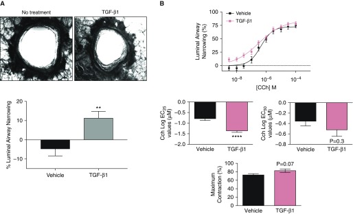
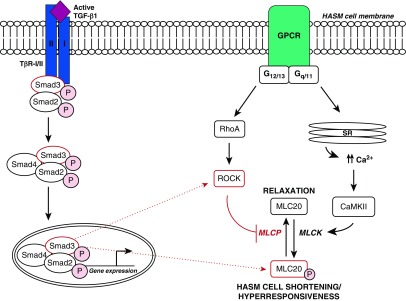
Transforming growth factor β1 (TGF-β1), a cytokine whose levels are elevated in the airways of patients with asthma, perpetuates airway inflammation and modulates airway structural cell remodeling. However, the role of TGF-β1 in excessive airway narrowing in asthma, or airway hyperresponsiveness (AHR), remains unclear. In this study, we set out to investigate the direct effects of TGF-β1 on human airway smooth muscle (HASM) cell shortening and hyperresponsiveness. The dynamics of AHR and single-cell excitation-contraction coupling were measured in human precision-cut lung slices and in isolated HASM cells using supravital microscopy and magnetic twisting cytometry, respectively. In human precision-cut lung slices, overnight treatment with TGF-β1 significantly augmented basal and carbachol-induced bronchoconstriction. In isolated HASM cells, TGF-β1 increased basal and methacholine-induced cytoskeletal stiffness in a dose- and time-dependent manner. TGF-β1-induced single-cell contraction was corroborated by concomitant increases in myosin light chain and myosin phosphatase target subunit 1 phosphorylation levels, which were attenuated by small interfering RNA-mediated knockdown of Smad3 and pharmacological inhibition of Rho kinase. Strikingly, these physiological effects of TGF-β1 occurred through a RhoA-independent mechanism, with little effect on HASM cell [Ca2+]i levels. Together, our data suggest that TGF-β1 enhances HASM excitation-contraction coupling pathways to induce HASM cell shortening and hyperresponsiveness. These findings reveal a potential link between airway injury-repair responses and bronchial hyperreactivity in asthma, and define TGF-β1 signaling as a potential target to reduce AHR in asthma.

Keywords: asthma; bronchoconstriction; contraction; cytokines; remodeling.


Figures

Figure 1.
Figure 1.
Airway narrowing in transforming growth factor β1 (TGF-β1)–treated human precision-cut lung slices (hPCLS). (A) Top: images were taken both before and after 18 hours of TGF-β1 exposure in one lung slice. Bottom: luminal airway narrowing in TGF-β1 (100 ng/mL, 18 h) and vehicle-treated hPCLS (n = 9 donors). (B) Carbachol (Cch)-induced luminal airway narrowing in TGF-β1 (100 ng/mL, 18 h) and vehicle-treated hPCLS (n = 9 donors). **P ≤ 0.01; ****P ≤ 0.0001. EC25 = 25% maximal effective concentration; EC50 = half-maximal effective concentration.
Figure 2.
Figure 2.
Agonist-induced human airway smooth muscle (HASM) cell [Ca2+]i and contractile responses of isolated HASM cells. (A) TGF-β1 (10–100 ng/mL) treatment increases the basal stiffness of isolated HASM cells (n = 221–477 individual cells). (B) SB-431542 (10 μM, 1 h pretreatment) decreases TGF-β1 (10 ng/mL, 18 h)-induced augmentation of basal HASM cell stiffness (n = 187–292). (C) SB-431542 (10 μM, 1 h pretreatment) decreases TGF-β1 (10 ng/mL, 18 h)-induced augmentation of Cch (n = 552–818) and histamine (Hist; n = 205–292 individual cells)–induced HASM cell stiffness. All magnetic twisting cytometry (MTC) data are represented as geometric mean (95% confidence interval). MTC statistical analysis was performed using one-way ANOVA with Tukey’s post hoc test. (D) Overnight TGF-β1 (10 ng/mL, 18 h) pretreatment augments [Ca2+]i in Cch-stimulated (10 μM) HASM cells. Three donors, two replicates per condition. *P ≤ 0.05; **P ≤ 0.01; ***P ≤ 0.001; ****P ≤ 0.0001. AUC = area under the curve; [Ca2+]i = intracellular calcium; ns = not significant; Pa = pascal.
Figure 3.
Figure 3.
MLC20 phosphorylation in TGF-β1–treated HASM cells. (A) TGF-β1 (10 ng/mL) induces time-dependent MLC20 phosphorylation (pMLC) in HASM cells (n = 3 ± SEM). (B) TGF-β1 (10 ng/mL) pretreatment for 18 hours augments Cch (10 μM, 10 min) and Hist–induced (1 μM, 10 min) MLC20 phosphorylation (n = 5; *P ≤ 0.05, ****P ≤ 0.0001). (C) Inhibition of TGF-β receptor I (TβR-I) signaling decreases TGF-β1–induced pMLC. For these studies, HASM cells were stimulated with TGF-β1 for a total of 18 hours. Prior to collection, HASM cells were treated with the ALK5 inhibitor SB-431542 (5 μM) for 1 hour, stimulated acutely with vehicle, Cch (10 μM), or Hist (1 μM), then lysed and subjected to immunoblot (n = 5 ± SEM). *P ≤ 0.05; **P < 0.01; ***P < 0.001; ****P ≤ 0.0001. MLC20 = 20 kD myosin light chain 20.
Figure 4.
Figure 4.
Effect of Rho kinase (ROCK) inhibition on TGF-β1–induced MLC20 phosphorylation and hyperresponsiveness in HASM cells. (A) HASM cells were treated with TGF-β1 (10 ng/mL) for 18 hours and then stimulated with Cch (10 μM) or Hist (1 μM) for 10 minutes (n = 5 ± SEM). (B) HASM cells were pretreated with the ROCK inhibitor Y-27632 for 15 minutes. HASM cells were then treated with TGF-β1 (10 ng/mL) for 18 hours and stimulated with Cch or Hist (n = 8 ± SEM, n = 4 ± SEM). pMYPT1/MYPT1 vehicle versus Y-27632 immunoblots show two different experiments representative of multiple donor observations. pMLC/MLC vehicle versus Y-27632 immunoblots show one representative immunoblot from multiple donor observations. (C) HASM cells were transfected with siRNA targeted against RhoA or a nontargeting (NT) siRNA pool, and then treated with TGF-β1 (10 ng/mL, 18 h) and stimulated with Cch (20 μM, 10 min; n = 3 ± SEM). *P < 0.05; **P < 0.01; ***P < 0.001. MYPT1 = myosin phosphatase target subunit 1; pMYTP1 = phosphorylated myosin phosphatase target subunit 1.
Figure 5.
Figure 5.
Effect of Smad knockdown on TGF-β1–induced MLC20 phosphorylation and hyperresponsiveness in HASM cells. HASM cells were transfected with siRNA against (A) Smad3 (n = 4–6 ± SEM) or (B) Smad2 (n = 3 ± SEM), pretreated with TGF-β1 (10 ng/mL) or vehicle control for 18 hours, and then stimulated with Cch (20 μM, 10 min). After treatment, HASM cells were lysed and subjected to immunoblot. **P < 0.01; ***P < 0.001.
Figure 6.
Figure 6.
TGF-β ligand and receptor basal gene expression in HASM cells derived from donors with no asthma (NA) or fatal asthma (FA). The mRNA expression of TGF-β (A) family ligands and (B) receptors was assessed by RNA sequencing in NA (n = 10) and FA (n = 5) donor–derived HASM cells. Shown are the transcripts with the highest expression levels, expressed as transcripts per million, for each gene.
Figure 7.
Figure 7.
Proposed role of TGF-β1 signaling in HASM cell shortening and hyperresponsiveness. After activated TGF-β1 binding to TβR-I/II, TβR-I kinase activity induces the phosphorylation of Smad2/3. After association with Smad4, the phosphorylated Smad2/3 complex translocates to the nucleus to mediate gene expression. Through a Smad3-dependent pathway, TGF-β1 augments HASM cell MLC phosphorylation and hyperresponsiveness to multiple agonists. Additionally, TGF-β1 exerts these effects through a ROCK-dependent, RhoA-independent pathway. However, further details regarding the mechanism by which Smad3 may induce ROCK, MLC phosphorylation, and hyperresponsiveness in HASM cells remain unclear. The dotted lines represent potential mechanisms regulating TGF-β1–induced HASM cell shortening and hyperresponsiveness. The figure is modified from a previously published image (6). Ca2+ = intracellular calcium; Ca2+/CaMKII = calcium/calmodulin-dependent kinase II; GPCR = G protein–coupled receptor; MLCK = myosin light light-chain kinase; MLCP = myosin light-chain phosphatase; SR = sarcoplasmic reticulum.

Comment in

References

    1. McDonald VM, Maltby S, Reddel HK, King GG, Wark PAB, Smith L, et al. Severe asthma: current management, targeted therapies and future directions—a roundtable report. 2017;22:53–60. - PubMed
    1. Carr TF, Bleecker E. Asthma heterogeneity and severity. 2016;9:41. - PMC - PubMed
    1. Hekking P-PW, Wener RR, Amelink M, Zwinderman AH, Bouvy ML, Bel EH. The prevalence of severe refractory asthma. 2015;135:896–902. - PubMed
    1. Sheehan WJ, Phipatanakul W. Difficult-to-control asthma: epidemiology and its link with environmental factors. 2015;15:397–401. - PMC - PubMed
    1. Yang Y, Zhang N, Lan F, Van Crombruggen K, Fang L, Hu G, et al. Transforming growth factor-β1 pathways in inflammatory airway diseases. 2014;69:699–707. - PubMed

Publication types

MeSH terms