

Utility of Molecular and Structural Brain Imaging to Predict Progression from Mild Cognitive Impairment to Dementia
- PMID: 28984586
- PMCID: PMC5679746
- DOI: 10.3233/JAD-161284
Utility of Molecular and Structural Brain Imaging to Predict Progression from Mild Cognitive Impairment to Dementia
Abstract
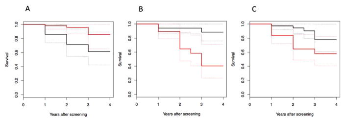
This project compares three neuroimaging biomarkers to predict progression to dementia in subjects with mild cognitive impairment (MCI). Eighty-eight subjects with MCI and 40 healthy controls (HCs) were recruited. Subjects had a 3T magnetic resonance imaging (MRI) scan, and two positron emission tomography (PET) scans, one with Pittsburgh compound B ([11C]PIB) and one with fluorodeoxyglucose ([18F]FDG). MCI subjects were followed for up to 4 y and progression to dementia was assessed on an annual basis. MCI subjects had higher [11C]PIB binding potential (BPND) than HCs in multiple brain regions, and lower hippocampus volumes. [11C]PIB BPND, [18F]FDG standard uptake value ratio (SUVR), and hippocampus volume were associated with time to progression to dementia using a Cox proportional hazards model. [18F]FDG SUVR demonstrated the most statistically significant association with progression, followed by [11C]PIB BPND and then hippocampus volume. [11C]PIB BPND and [18F]FDG SUVR were independently predictive, suggesting that combining these measures is useful to increase accuracy in the prediction of progression to dementia. Hippocampus volume also had independent predictive properties to [11C]PIB BPND, but did not add predictive power when combined with the [18F]FDG SUVR data. This work suggests that PET imaging with both [11C]PIB and [18F]FDG may help to determine which MCI subjects are likely to progress to AD, possibly directing future treatment options.
Keywords: Alzheimer’s disease; PET; mild cognitive impairment; prognosis; volumetric MRI.
Conflict of interest statement
Figures
References
-
- Scheltens P, Blennow K, Breteler MM, de Strooper B, Frisoni GB, Salloway S, Van der Flier WM. Alzheimer’s disease. Lancet 2016 - PubMed
-
- Sperling RA, Aisen PS, Beckett LA, Bennett DA, Craft S, Fagan AM, Iwatsubo T, Jack CR, Jr, Kaye J, Montine TJ, Park DC, Reiman EM, Rowe CC, Siemers E, Stern Y, Yaffe K, Carrillo MC, Thies B, Morrison-Bogorad M, Wagster MV, Phelps CH. Toward defining the preclinical stages of Alzheimer’s disease: recommendations from the National Institute on Aging-Alzheimer’s Association workgroups on diagnostic guidelines for Alzheimer’s disease. Alzheimers Dement. 2011;7:280–292. - PMC - PubMed
-
- He W, Liu D, Radua J, Li G, Han B, Sun Z. Meta-analytic comparison between PIB-PET and FDG-PET results in Alzheimer’s disease and MCI. Cell Biochem Biophys. 2015;71:17–26. - PubMed
-
- Devanand DP, Pradhaban G, Liu X, Khandji A, De Santi S, Segal S, Rusinek H, Pelton GH, Honig LS, Mayeux R, Stern Y, Tabert MH, de Leon MJ. Hippocampal and entorhinal atrophy in mild cognitive impairment: prediction of Alzheimer disease. Neurology. 2007;68:828–836. - PubMed
MeSH terms
Substances
Grants and funding
LinkOut - more resources
Full Text Sources
Other Literature Sources
Medical
