. 2018 Feb;191(2):151-165.
doi: 10.1111/cei.13066. Epub 2017 Nov 20.

B cell tolerance to epidermal ribonuclear-associated neo-autoantigen in vivo

Affiliations

B cell tolerance to epidermal ribonuclear-associated neo-autoantigen in vivo

S E Degn et al. Clin Exp Immunol. 2018 Feb.

Abstract

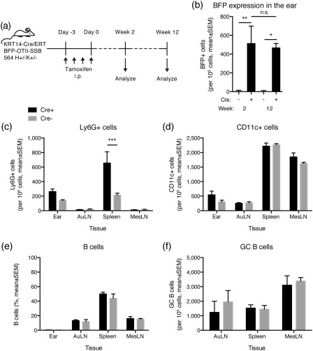
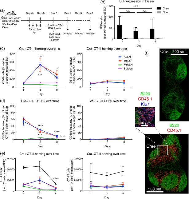
Defining how self-antigens are perceived by the immune system is pivotal to understand how tolerance is maintained under homeostatic conditions. Clinically relevant, natural autoantigens targeted by autoantibodies, in e.g. systemic lupus erythematosus (SLE), commonly have an intrinsic ability to engage not only the B cell receptor (BCR), but also a co-stimulatory pathway in B cells, such as the Toll-like receptor (TLR)-7 pathway. Here we developed a novel mouse model displaying inducible expression of a fluorescent epidermal neo-autoantigen carrying an OT-II T cell epitope, B cell antigen and associated ribonucleic acids capable of stimulating TLR-7. The neo-autoantigen was expressed in skin, but did not drain in intact form into draining lymph nodes, even after ultraviolet B (UVB)-stimulated induction of apoptosis in the basal layer. Adoptively transferred autoreactive B cells were excluded follicularly and perished at the T-B border in the spleen, preventing their recirculation and encounter with antigen peripherally. This transitional check-point was bypassed by crossing the reporter to a BCR knock-in line on a C4-deficient background. Adoptively transferred OT-II T cells homed rapidly into cutaneous lymph nodes and up-regulated CD69. Surprisingly, however, tolerance was not broken, as the T cells subsequently down-regulated activation markers and contracted. Our results highlight how sequestration of intracellular and peripheral antigen, the transitional B cell tolerance check-point and T cell regulation co-operate to maintain immunological tolerance in vivo.

Keywords: B cell; autoimmunity; skin antigen; systemic lupus erythematosus; tolerance.


Figures

Figure 1
Figure 1
Generation of blue fluorescent protein‐OTII epitope‐Sjögren's syndrome B protein (BFP‐OTII‐SSB) reporter. (a) Schematic overview of the targeting vector. A CAG promotor is situated upstream of a floxed stop region, followed by the open reading frame for BFP, the OTII peptide sequence and SSB protein. This is followed by a woodchuck hepatitis virus post‐transcriptional regulatory element (WPRE) and a bovine growth hormone polyadenylation signal (BGH‐pA), and finally a phosphoglycerate kinase promotor (PGK) driving expression of the neomycin selection marker. The full cassette is flanked by targeting arms for stable integration into the Rosa26 locus, and a Diphtheria toxin A fragment expression cassette (PGK‐DTA‐BGH pA) drives negative selection of random integration events. (b) Southern blot of HindIII digested genomic DNA extracted from transfected BRUCE 4 embryonic stem (ES) cells after neomycin selection. Results for six different ES clones, labelled 1–6, are shown. Expected wild‐type band is 4·5 kb, whereas the mutant targeted allele is 5·6 kb. As can be seen, two of six ES clones harbour the knock‐in. (c) Example of genotyping results for the knock‐in line. Top gel shows polymerase chain reaction (PCR) for wild‐type, for seven unknown tail DNA samples (1–7), a B6 control and a heterozygous knock‐in control. Bottom gel shows PCR for knock‐in allele for the same samples.
Figure 2
Figure 2
Tamoxifen dependence of blue fluorescent protein‐OTII epitope‐Sjögren's syndrome B protein (BFP‐OTII‐SSB) expression, subcellular and gross histological localization. (a) Schematic overview of the knock‐in BFP‐OTII‐SSB Rosa locus and the tamoxifen‐induced Cre recombination event permitting its expression. Tamoxifen‐controlled nuclear translocation of Cre expressed under the KRT14 promotor allows Cre‐mediated excision of the stop cassette blocking expression of BFP‐OTII‐SSB fusion protein. (b) Representative images of ear skin either untreated (left panels) or after tamoxifen treatment regimen (right panels). Blue = BFP; red = phalloidin‐A568 counterstain. Scale bar indicated. The two images for the untreated sample are representative of either end of the spectrum observed, i.e. no background (left, top image) and the highest level of observed background (left, bottom image). (c) Quantification of mean fluorescence intensity (MFI) in ear skin of untreated (–) versus tamoxifen‐treated (+) animals. Total image MFI was measured in ImageJ. Individual data points and mean ± standard deviation (s.d.) for two slices per stack for three to four stacks per animal, for two mice in each group, from two independent experiments. Statistical significance given for two‐tailed unpaired Mann–Whitney test. (d) Subcellular localization of BFP signal. Top: transection of epidermis, showing nuclear localization of BFP, counterstained with phalloidin in red. Inset: high magnification view of nuclear and nucleolar localized BFP in the epidermis, with nuclear counterstain [7‐aminoactinomycin D (7AAD)] in red. Bottom: ultra‐high magnification image showing defined nucleolar and diffuse nuclear localization of BFP in relation to nuclear counterstain (7AAD) in red. (e) Left: low magnification overview of BFP signal in transection of the ear, with overlay of the differential interference contrast (DIC) channel for gross morphology. Right: low magnification overview of BFP signal in transection of the ear, with overlay of propidium iodide (PI) for nuclear counterstain. Top panels are from Cre mice, bottom panels are from Cre+ mice.
Figure 3
Figure 3
Establishment of ultraviolet B (UVB) irradiation protocol. (a) A UVB irradiation protocol was established allowing timing of induction of apoptosis in the basal layer of ear skin, as evidenced by cleaved caspase 3 positivity in the basal layer at day 1 post‐irradiation (top, left panel), gross enlargement of the draining auricular lymph node compared to the contralateral control (top, middle panel), influx of Ly6G+ neutrophils into the medulla of the node (top, right panel), expansion of the Lyve‐1+ lymphatic network and induction of B cell clusters in the medulla (bottom panel). Images are representative of observations in more than five mice. (b) Quantification of cleaved caspase 3 signal in the basal layer of ear skin. (c) Quantification of BFP signal in the basal layer. For b and c, n = 15 measurements from five mice for the non‐irradiated group and n = 16 measurements from five mice for the irradiated group, pooled from two independent experiments. (d) Quantification of Ly6G+ infiltrating cells in the ear, measured in a subset of the samples presented in b and c. (e) Quantification of CD207+ Langerhans cells in the ear, for a subset of the samples presented in b and c. In b–e, lines represent mean ± standard error of the mean (s.e.m.). The exact P‐values for two‐tailed Mann–Whitney comparison of non‐irradiated and irradiated groups are given for each of the parameters in their respective graph.
Figure 4
Figure 4
Adoptively transferred OT‐II T cells home to skin‐draining lymph nodes and up‐regulate CD69, whereas the bulk of adoptively transferred 564Igi B cells are follicularly excluded in spleen and eliminated. (a) Overview of experimental layout. Mice were used that carry a KRT14‐driven Cre under the control of a mutated form of the ligand‐binding domain of the oestrogen receptor that only binds tamoxifen (ERT), and a floxed‐stop expression cassette for a fusion construct of blue fluorescent protein‐OTII epitope‐Sjögren's syndrome B protein (BFP‐OTII‐SSB). Expression of BFP‐OTII‐SSB was turned on in the skin by intraperitoneal (i.p.) administration of tamoxifen (days −6 to −3), followed by ultraviolet B (UVB) irradiation of both ears to induce apoptosis in the basal layer (day −1). Subsequently, 5 × 106 OT‐II CD4 T cells and 1 × 107 564Igi B cells were transferred adoptively into the mice by intravenous (i.v.) injection (day 0). Spleen and auricular lymph nodes were analysed on day 1 or day 3. (b) Adoptively transferred OT‐II cells home preferentially to skin‐draining lymph nodes. (c) Adoptively transferred OT‐II cells up‐regulate CD69 in skin‐draining lymph nodes at day 1, then down‐regulate it again by day 3. (d) On day 1, adoptively transferred 564Igi B cells remain mainly in spleen due to follicular exclusion, and few escape to peripheral skin‐draining nodes. By day 3, the majority of follicularly excluded 564Igi B cells have perished. For b–d, mean ± standard error of the mean (s.e.m.) is indicated. For control spleen, n = 1, whereas for spleen and AuLN, n = 2, with each point representative of two mice (samples pooled 2 and 2), i.e. n = 4 per time‐point for a total number of eight animals. P‐values given for regular two‐way analysis of variance (anova) with Sidak's post‐test. Representative of two experiments with similar results. (e) Serial sections from a representative spleen sample illustrating follicular exclusion of adoptively transferred carboxyfluorescein succinimidyl ester (CFSE)‐labelled 564Igi B cells, as demonstrated by counterstaining for: CD3 in blue and B220 in red (top); immunoglobulin (Ig)D in blue and IgM in red (middle); CD21 in blue and CD169 in red (bottom).
Figure 5
Figure 5
Long‐term consequences of blue fluorescent protein‐OTII epitope‐Sjögren's syndrome B protein (BFP‐OTII‐SSB) expression. (a) Overview of experimental layout. 564Igi mice were crossed with mice that carry a KRT14‐driven Cre under the control of a mutated form of the ligand‐binding domain of the oestrogen receptor that only binds tamoxifen (ERT), and a floxed‐stop expression cassette for a fusion construct of BFP‐OTII‐SSB, and Cre and Cre+ littermates were used. Expression of BFP‐OTII‐SSB was turned on in the skin by intraperitoneal (i.p.) administration of tamoxifen (days −3 to 0). The ear skin, auricular lymph nodes, spleen and mesenteric lymph nodes were analysed at 2 or 12 weeks. (b) BFP expression in ears of KRT14 Cre (grey bars) versus Cre+ (black bars) BFP‐OTII‐SSB 564 H+/– K+/– mice at 2 and 12 weeks after tamoxifen application. Mean ± standard error of the mean (s.e.m.) is shown. For week 2 measurements, n = 6 in each group, for week 12 measurements, n = 4 for Cre+ and n = 3 for Cre. Cre+ and Cre were compared by two‐way analysis of variance (anova) with Holm–Sidak's post‐test (*P < 0·05; **P < 0·01). There was no statistically significant difference between the time‐points for either Cre or Cre+, as assessed by two‐way anova with Holm–Sidak's post‐test (n.s. = not significant). (c) Frequency of Ly6G‐positive cells in ear, auricular lymph nodes, spleen and mesenteric lymph nodes of Cre+ (black bars, mean ± s.e.m. of n = 4 mice) versus Cre (grey bars, mean ± s.e.m. of n = 3 mice) at 12 weeks post‐tamoxifen treatment. Statistical significance of differences between Cre+ and Cre assessed by two‐way anova with Sidak's multiple comparisons test (***P < 0·001). (d) As c, but showing CD11c+ cell frequencies. (e) As c, but showing B cell frequencies. (f) As c, but showing GC B cell frequencies.
Figure 6
Figure 6
Adoptively transferred OT‐II T cells home to skin‐draining lymph nodes, independent of ultraviolet B (UVB) irradiation, show hallmarks of activation and expand then subsequently contract again. (a) Schematic overview of experimental layout. 564Igi C4–/– mice were crossed with mice that carry a KRT14‐driven Cre under the control of a mutated form of the ligand‐binding domain of the oestrogen receptor that only binds tamoxifen (ERT), and a floxed‐stop expression cassette for a fusion construct of blue fluorescent protein, an OTII epitope, and Sjögren's syndrome B protein (BFP‐OTII‐SSB), on a C4‐deficient background. Cre and Cre+ littermates were used, and expression of BFP‐OTII‐SSB was turned on in the skin by intraperitoneal (i.p.) administration of tamoxifen (days −6 to −3). This was followed by UVB irradiation of both ears to induce apoptosis in the basal layer, and adoptive transfer of 1 × 107 OT‐II CD4 T cells by intravenous (i.v.) injection (day 0). Ear skin, auricular lymph nodes, inguinal lymph nodes, mesenteric lymph nodes and spleen were analyzed on days 1, 3 or 8. (b) BFP expression in the ears of adoptive transfer recipients. Two‐way analysis of variance (anova) with Sidak's multiple comparisons test (n.s. = not significant). (c) Distribution of OT‐II cells (as percent relative to spleen) following adoptive transfer into UVB irradiated KRT14 Cre+ (left panel) versus Cre (right panel) BFP‐OTII‐SSB 564Igi H+/–K+/– C4–/– recipients. (d) CD69 expression on adoptively transferred OT‐II cells. (e) Absolute frequencies of OT‐II cells following adoptive transfer. For b–e, mean ± standard error of the mean (s.e.m.) of measurements from two mice are shown per group, per time‐point, for a total of 12 mice. Results are representative of two experiments with similar results. For c–e, statistical significance is indicated for two‐way anova with Dunnett's multiple comparisons test (*P < 0·05; ***P < 0·001; ****P < 0·0001). (f) Stitched whole‐node views of inguinal lymph node from a Cre (top) or Cre+ (bottom) mouse at day 3. B220 in green, CD45.1+ OT‐II T cells in red. Scale bar indicated next to the top image. The blow‐out from the Cre+ node is at an additional ×2 magnification, and includes Ki67 staining in blue.

Similar articles

Cited by

References

    1. Fiorina P, Sayegh MH. B cell‐targeted therapies in autoimmunity: rationale and progress. F1000 Biol Rep 2009; 1:39. - PMC - PubMed
    1. Kleffel S, Vergani A, Tezza S et al Interleukin‐10+ regulatory B cells arise within antigen‐experienced CD40+ B cells to maintain tolerance to islet autoantigens. Diabetes 2015; 64:158–71. - PMC - PubMed
    1. Fiorina P, Vergani A, Dada S et al Targeting CD22 reprograms B‐cells and reverses autoimmune diabetes. Diabetes 2008; 57:3013–24. - PMC - PubMed
    1. Matzinger P. Tolerance, danger, and the extended family. Annu Rev Immunol 1994; 12:991–1045. - PubMed
    1. Berland R, Fernandez L, Kari E et al Toll‐like receptor 7‐dependent loss of B cell tolerance in pathogenic autoantibody knockin mice. Immunity 2006; 25:429–40. - PubMed

Publication types

MeSH terms