Review
. 2018 Jan;25(1):10.1111/micc.12421.
doi: 10.1111/micc.12421.

KV channels and the regulation of vascular smooth muscle tone

Affiliations
Review

KV channels and the regulation of vascular smooth muscle tone

William F Jackson. Microcirculation. 2018 Jan.

Abstract

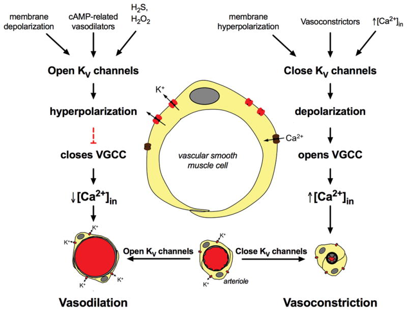
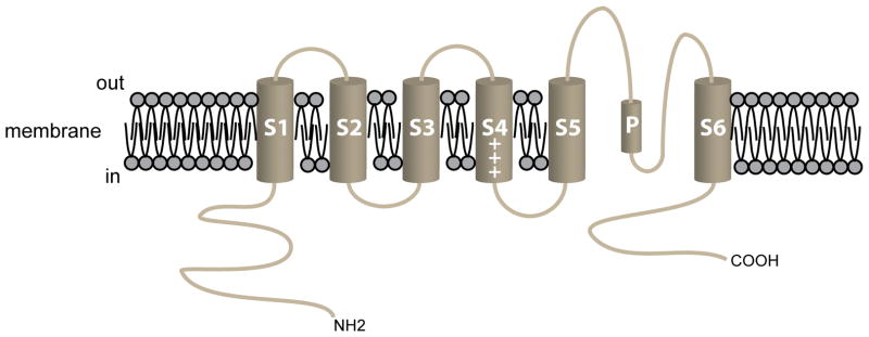
VSMCs in resistance arteries and arterioles express a diverse array of KV channels with members of the KV 1, KV 2 and KV 7 families being particularly important. Members of the KV channel family: (i) are highly expressed in VSMCs; (ii) are active at the resting membrane potential of VSMCs in vivo (-45 to -30 mV); (iii) contribute to the negative feedback regulation of VSMC membrane potential and myogenic tone; (iv) are activated by cAMP-related vasodilators, hydrogen sulfide and hydrogen peroxide; (v) are inhibited by increases in intracellular Ca2+ and vasoconstrictors that signal through Gq -coupled receptors; (vi) are involved in the proliferative phenotype of VSMCs; and (vii) are modulated by diseases such as hypertension, obesity, the metabolic syndrome and diabetes. Thus, KV channels participate in every aspect of the regulation of VSMC function in both health and disease.

Keywords: KV channels; arterioles; blood flow; microcirculation; potassium channels; resistance arteries; vascular smooth muscle; vasoconstriction; vasodilation.


Figures

Figure 1
Figure 1
KV channels and the regulation of arteriolar tone. Left side of the diagram shows several factors that activate KV channels in VSMCs (see text for more information and references) leading to K+ -efflux through these channels, membrane hyperpolarization and ultimately, vasodilation. Right side of diagram shows factors that inhibit KV channel activity (see text for details and references), leading to membrane depolarization and vasoconstriction.
Figure 2
Figure 2
The pore-forming α-subunit of KV channels. Schematic diagram of the 6 membrane spanning domains of a typical KV channel. The length and composition of the amino (NH2) and the carboxy (COOH) termini vary among the large number of KV channel isoforms expressed in VSMCs. For KV1.5, the NH2-terminus in the full-length version of the channel is 247 amino acids (AAs) and the COOH-terminus is 89 AAs in length. In KV1.5, the selectivity filter (AAs 480–485) follows the 9 AA, pore helix (AAs 468–479), labeled “P” in the diagram. Modulatory KVβ-subunits interact with residues in the NH2-terminus of the α-subunit. Post-synaptic density (PSD)-95 interacts with PDZ domains in the COOH-terminus of KV channels.
Figure 3
Figure 3
KV channel block with 4-aminopyridine inhibits membrane K+ currents, depolarizes VSMCs and constricts arterioles. Panel A shows the family of outward K+ currents elicited by 10 mV depolarizing voltage steps from −90 mV to 0 mV recorded in hamster cremasteric arteriolar VSMCs using the perforated patch technique [90]. Panel B shows currents in the same cell after superfusion with 3 mM 4-aminopyridine (4-AP): outward currents are virtually abolished. Panel C shows summary means ± SEM for similar experiments showing the current-voltage relationship for the data shown in Panels A and B. Panel D shows the current-voltage relationship from Panel C on an expanded scale to show that 4-AP (3 mM) inhibits currents around the resting membrane potential of these cells (~-44 mV), and shifts the reversal potential for whole-cell currents (a measure of membrane potential) to more positive potentials. Panel E shows effects of 4-AP (3 mM) on membrane potential of an isolated hamster cremasteric arteriolar VSMC recorded in current-clamp experiments using the perforated-patch technique: 4-AP depolarized the cell from ~-40 mV to ~-25 mV, consistent with the data shown in Panel D. Panel F shows summary data for experiments as in Panel E demonstrating that 4-AP (3 mM) consistently depolarizes hamster cremasteric arteriolar VSMCs. Panel G shows data from pressure-myography experiments demonstrating that 4-AP consistently constricts isolated second-order cremasteric arterioles, consistent with VSMC depolarization (Panels D–F) due to block of outward KV currents (Panels A–C). These data show 4-AP-sensitive KV channel currents contribute to resting membrane potential and myogenic tone in arteriolar VSMCs. Data Panels A–D modified from [90], with permission. Data in Panels E–G replotted from [88], with permission.

Similar articles

Cited by

References

    1. Abbott GW, Jepps TA. Kcne4 Deletion Sex-Dependently Alters Vascular Reactivity. J Vasc Res. 2016;53:138–148. - PMC - PubMed
    1. Aiello EA, Clement-Chomienne O, Sontag DP, Walsh MP, Cole WC. Protein kinase C inhibits delayed rectifier K+ current in rabbit vascular smooth muscle cells. Am J Physiol. 1996;271:H109–119. - PubMed
    1. Aiello EA, Malcolm AT, Walsh MP, Cole WC. Beta-adrenoceptor activation and PKA regulate delayed rectifier K+ channels of vascular smooth muscle cells. American Journal of Physiology. 1998;275:H448–H459. - PubMed
    1. Aiello EA, Walsh MP, Cole WC. Isoproterenol and forskolin increase and PKI inhibits delayed rectifier K+ current in vascular myocytes isolated from rabbit coronary artery and portal vein. CanJPhysiolPharmacol. 1994;72:47.
    1. Aiello EA, Walsh MP, Cole WC. Phosphorylation by protein kinase A enhances delayed rectifier K+ current in rabbit vascular smooth muscle cells. AmJPhysiolHeart CircPhysiol. 1995;268:H926–H934. - PubMed

Publication types

Substances

LinkOut - more resources