. 2018 Jan:42:11-20.
doi: 10.1016/j.cellsig.2017.10.001. Epub 2017 Oct 3.

A cellular threshold for active ERK1/2 levels determines Raf/MEK/ERK-mediated growth arrest versus death responses

Affiliations

A cellular threshold for active ERK1/2 levels determines Raf/MEK/ERK-mediated growth arrest versus death responses

Seung-Keun Hong et al. Cell Signal. 2018 Jan.

Abstract

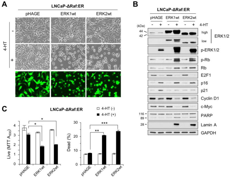
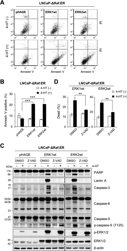
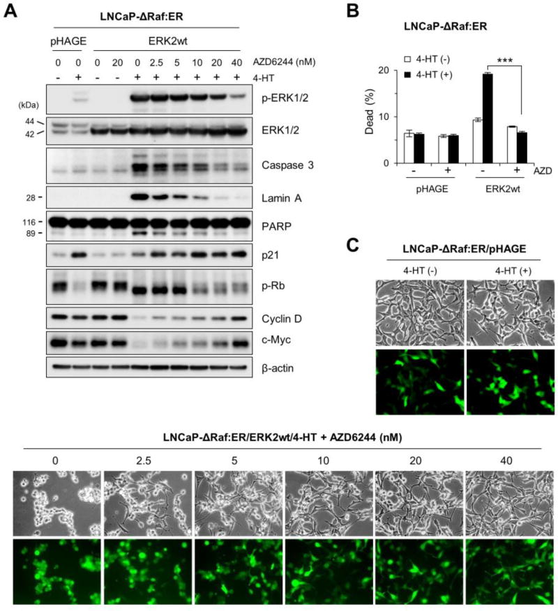
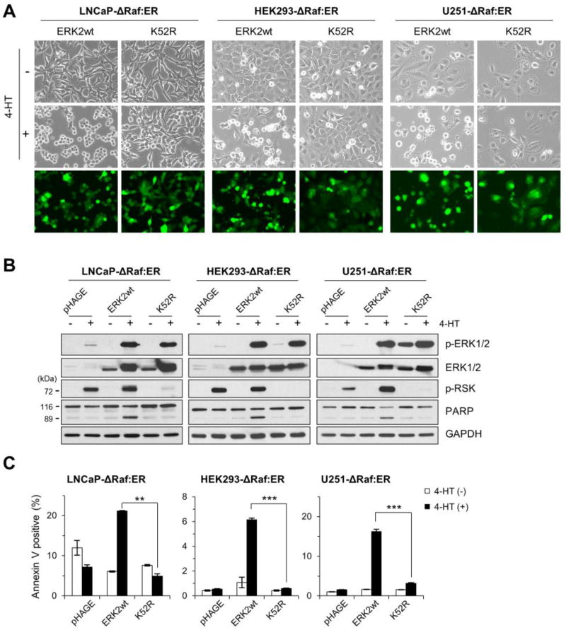
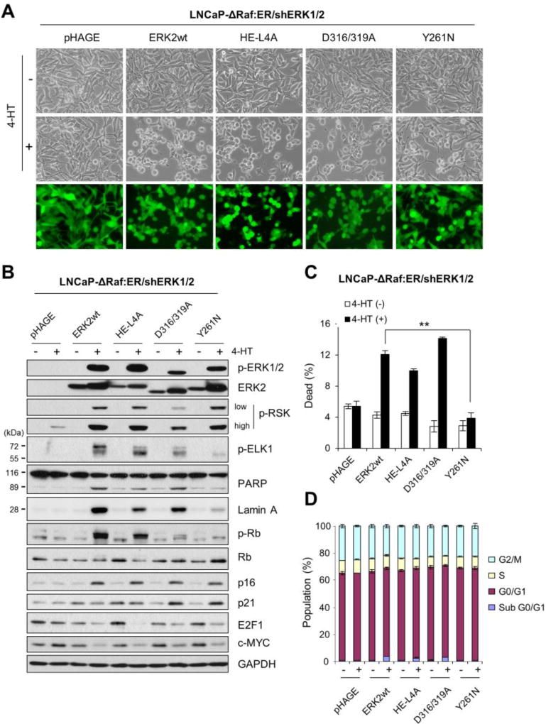
In addition to its conventional role for cell proliferation and survival, the Raf/MEK/Extracellular signal-regulated kinase (ERK) pathway can also induce growth arrest and death responses, if aberrantly activated. Here, we determined a molecular basis of ERK1/2 signaling that underlies these growth inhibitory physiological outputs. We found that overexpression of ERK1 or ERK2 switches ΔRaf-1:ER-induced growth arrest responses to caspase-dependent apoptotic death responses in different cell types. These death responses, however, were reverted to growth arrest responses upon titration of cellular phospho-ERK1/2 levels by the MEK1/2 inhibitor AZD6244. These data suggest that a cellular threshold for active ERK1/2 levels exists and affects the cell fate between death and growth arrest. We also found that death-mediating ability of ERK2 is abolished by the catalytic site-disabling Lys52Arg replacement or significantly attenuated by the F-site recruitment site-disabling Tyr261Asn replacement, although unaffected by the mutations that disable the common docking groove or the dimerization interface. Therefore, ERK1/2 mediates death signaling dependently of kinase activity and specific physical interactions. Intriguingly, Tyr261Asn-replaced ERK2 could still mediate growth arrest signaling, further contrasting the molecular basis of ERK1/2-mediated growth arrest and death signaling. These data reveal a mechanism underlying the role of ERK1/2 as a focal point of Raf/MEK/ERK-mediated growth arrest and death signaling.

Keywords: Cell death; ERK1/2; Growth arrest; MEK1/2; Raf.


Conflict of interest statement

Conflict of interest

The authors declare no conflict of interest for this article.

Figures

Fig 1
Fig 1. Overexpression of ERK1 or ERK2 switches ΔRaf-1:ER-induced growth arrest responses to death responses
LNCaP-ΔRaf-1:ER cells, infected with the lentiviral pHAGE-ERK1, pHAGE-ERK2, or the control pHAGE, were treated with 1 µM 4-hydroxytamoxifen (4-HT) for 24 hours. Cells were examined for morphological changes (A), expression of the indicated proteins by Western blot analysis (B), and viability by MTT assay and trypan blue exclusion analysis (C). Similar infection efficiency was verified by GFP expression (bottom panels in A). p-ERK1/2 indicates ERK1/2 phosphorylated at Thr202/Tyr204 (ERK1) and Thr183/Tyr185 (ERK2). p-Rb indicates Rb phosphorylated at Ser780. GAPDH is the control for equal protein loading. Data (means ± SE) are from a representative experiment performed in triplicate. *, P < 0.05; ***, P < 0.001.
Fig 2
Fig 2. Overexpression of ERK1 or ERK2 induces caspase-dependent apoptotic cell death in ΔRaf-1:ER-activated cells
(A and B) LNCaP-ΔRaf-1:ER cells, infected with the lentiviral pHAGE-ERK1, pHAGE-ERK2, or pHAGE, were treated with 1 µM 4-HT for 24 hours prior to annexin V/propidium iodide (PI) staining. The graphs (B) indicate annexin V-positive cell populations. (C and D) LNCaP-ΔRaf-1:ER cells, infected with the lentiviral pHAGE-ERK1, pHAGE-ERK2, or pHAGE, were treated with 1 µM 4-HT for 1 day in the presence of the pan-caspase inhibitor, Z-VAD(OMe)-FMK (20 µM). Cells were examined for expression of the indicated proteins by Western blot analysis (C) and viability by trypan blue exclusion analysis (D). Caspase-3 is cleaved into 17 and 19 kDa peptides. Caspase-8 (50/55 kDa doublets) is cleaved into 40/36 kDa doublets and 23 kDa peptides. Caspase-9 (47 kDa) is cleaved into 37/35 kDa peptides. β-actin is the control for equal protein loading. Data (mean ± SEM) are from a representative experiment performed in triplicate. **P < 0.01; ***P < 0.001.
Fig 3
Fig 3. AZD6244 reverts ERK2 overexpression-mediated cell death responses to growth arrest responses in ΔRaf-1:ER-activated cells
LNCaP-ΔRaf-1:ER cells, infected with the lentiviral pHAGE-ERK2 or pHAGE, were treated with 1 µM 4-HT for 24 hours in the presence of the MEK1/2 inhibitor AZD6244. Cells were examined for expression of the indicated proteins by Western blot analysis (A), viability by trypan blue exclusion analysis (B), and morphological changes (C). Similar infection efficiency was verified by GFP expression (bottom panels in C). Caspase-3 is cleaved into 17 and 19 kDa peptides. β-actin is the control for equal protein loading. Data (means ± SE) are from a representative experiment performed in triplicate. ***, P < 0.001.
Fig 4
Fig 4. Kinase-deficient ERK2 cannot mediate cell death signaling
LNCaP-ΔRaf-1:ER, HEK293-ΔRaf-1:ER, and U251-ΔRaf-1:ER cells, infected with the lentiviral pHAGE-ERK2, pHAGE-ERK2-K52R, or pHAGE, were treated with 1 µM 4-HT for 24 hours. Cells were then examined for morphological changes (A), expression of the indicated proteins by Western blot analysis (B), and apoptosis by annexin V/propidium iodide staining (C). Similar infection efficiency was verified by GFP expression (bottom panels in A). p-RSK indicates p90RSK phosphorylated at Thr359/Ser363. GAPDH is the control for equal protein loading. Data (means ± SE) are from a representative experiment performed in triplicate. **, P < 0.01; ***, P < 0.001.
Fig 5
Fig 5. FRS-disabled ERK2 cannot mediate cell death signaling
LNCaP-ΔRaf-1:ER/shERK1/2 cells, infected with the lentiviral pHAGE-ERK2, pHAGE-ERK2-HE/L4A, pHAGE-ERK2-D316/319A, pHAGE-ERK2-Y261N, or pHAGE, were treated with 1 µM 4-HT for 24 hours. Cells were examined for morphological changes (A), expression of the indicated proteins by Western blot analysis (B), viability by trypan blue exclusion analysis (C), and cell cycle analysis (D). Similar infection efficiency was verified by GFP expression (bottom panels in A). p-ELK1 indicates ELK1 phosphorylated at Ser383. GAPDH is the control for equal protein loading. Data (means ± SE) are from a representative experiment performed in triplicate. **, P < 0.01.

References

    1. Roskoski R., Jr ERK1/2 MAP kinases: structure, function, and regulation. Pharmacol Res. 2012;66:105–143. - PubMed
    1. Shaul YD, Seger R. The MEK/ERK cascade: from signaling specificity to diverse functions. Biochim Biophys Acta. 2007;1773:1213–1226. - PubMed
    1. Chambard JC, Lefloch R, Pouyssegur J, Lenormand P. ERK implication in cell cycle regulation. Biochim Biophys Acta. 2007;1773:1299–1310. - PubMed
    1. Balmanno K, Cook SJ. Tumour cell survival signalling by the ERK1/2 pathway. Cell death and differentiation. 2009;16:368–377. - PubMed
    1. McCubrey JA, Steelman LS, Chappell WH, Abrams SL, Montalto G, Cervello M, Nicoletti F, Fagone P, Malaponte G, Mazzarino MC, Candido S, Libra M, Basecke J, Mijatovic S, Maksimovic-Ivanic D, Milella M, Tafuri A, Cocco L, Evangelisti C, Chiarini F, Martelli AM. Mutations and deregulation of Ras/Raf/MEK/ERK and PI3K/PTEN/Akt/mTOR cascades which alter therapy response. Oncotarget. 2012;3:954–987. - PMC - PubMed

MeSH terms