

Brain Activity Unique to Orgasm in Women: An fMRI Analysis
- PMID: 28986148
- PMCID: PMC5675825
- DOI: 10.1016/j.jsxm.2017.08.014
Brain Activity Unique to Orgasm in Women: An fMRI Analysis
Abstract
Background: Although the literature on imaging of regional brain activity during sexual arousal in women and men is extensive and largely consistent, that on orgasm is relatively limited and variable, owing in part to the methodologic challenges posed by variability in latency to orgasm in participants and head movement.
Aim: To compare brain activity at orgasm (self- and partner-induced) with that at the onset of genital stimulation, immediately before the onset of orgasm, and immediately after the cessation of orgasm and to upgrade the methodology for obtaining and analyzing functional magnetic resonance imaging (fMRI) findings.
Methods: Using fMRI, we sampled equivalent time points across female participants' variable durations of stimulation and orgasm in response to self- and partner-induced clitoral stimulation. The first 20-second epoch of orgasm was contrasted with the 20-second epochs at the beginning of stimulation and immediately before and after orgasm. Separate analyses were conducted for whole-brain and brainstem regions of interest. For a finer-grained analysis of the peri-orgasm phase, we conducted a time-course analysis on regions of interest. Head movement was minimized to a mean less than 1.3 mm using a custom-fitted thermoplastic whole-head and neck brace stabilizer.
Outcomes: Ten women experienced orgasm elicited by self- and partner-induced genital stimulation in a Siemens 3-T Trio fMRI scanner.
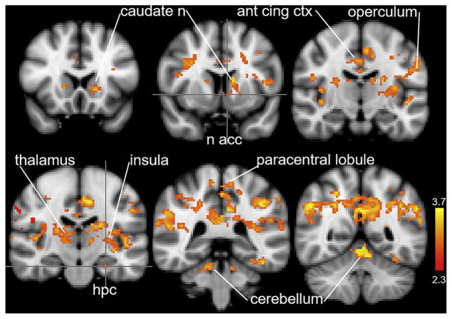
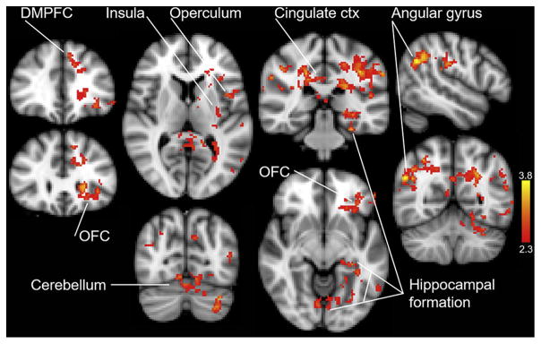
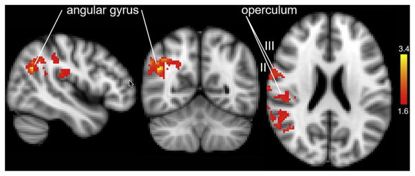
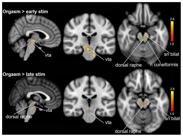
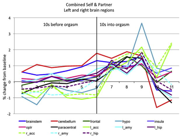
Results: Brain activity gradually increased leading up to orgasm, peaked at orgasm, and then decreased. We found no evidence of deactivation of brain regions leading up to or during orgasm. The activated brain regions included sensory, motor, reward, frontal cortical, and brainstem regions (eg, nucleus accumbens, insula, anterior cingulate cortex, orbitofrontal cortex, operculum, right angular gyrus, paracentral lobule, cerebellum, hippocampus, amygdala, hypothalamus, ventral tegmental area, and dorsal raphe).
Clinical translation: Insight gained from the present findings could provide guidance toward a rational basis for treatment of orgasmic disorders, including anorgasmia.
Strengths and limitations: This is evidently the first fMRI study of orgasm elicited by self- and partner-induced genital stimulation in women. Methodologic solutions to the technical issues posed by excessive head movement and variable latencies to orgasm were successfully applied in the present study, enabling identification of brain regions involved in orgasm. Limitations include the small sample (N = 10), which combined self- and partner-induced stimulation datasets for analysis and which qualify the generalization of our conclusions.
Conclusion: Extensive cortical, subcortical, and brainstem regions reach peak levels of activity at orgasm. Wise NJ, Frangos E, Komisaruk BR. Brain Activity Unique to Orgasm in Women: An fMRI Analysis. J Sex Med 2017;14:1380-1391.
Keywords: Functional Magnetic Resonance Imaging; Human Female; Orgasm; Sexual Arousal; Sexual Behavior.
Copyright © 2017 International Society for Sexual Medicine. Published by Elsevier Inc. All rights reserved.
Conflict of interest statement
Figures
References
-
- Hamann S, Herman RA, Nolan CL, et al. Men and women differ in amygdala response to visual sexual stimuli. Nat Neurosci. 2004;7:411–416. - PubMed
-
- Feretti A, Caulo M, Del Gratta C, et al. Dynamics of male sexual arousal: distinct components of brain activation revealed by fMRI. Neuroimage. 2005;15:1086–1096. - PubMed
Publication types
MeSH terms
Grants and funding
LinkOut - more resources
Full Text Sources
Other Literature Sources
