. 2017 Dec 1;292(48):19693-19707.
doi: 10.1074/jbc.RA117.000130. Epub 2017 Oct 6.

The SecA protein deeply penetrates into the SecYEG channel during insertion, contacting most channel transmembrane helices and periplasmic regions

Affiliations

The SecA protein deeply penetrates into the SecYEG channel during insertion, contacting most channel transmembrane helices and periplasmic regions

Tithi Banerjee et al. J Biol Chem. .

Abstract

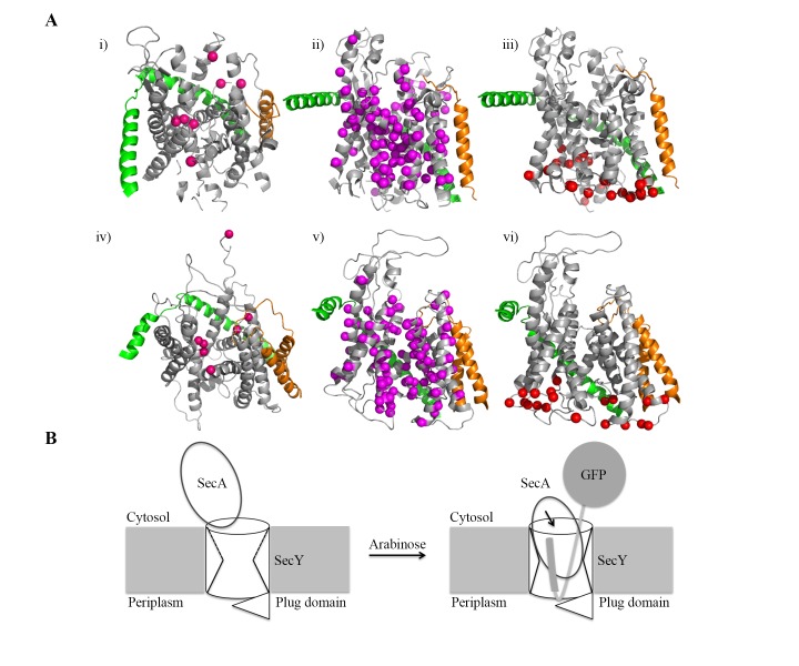
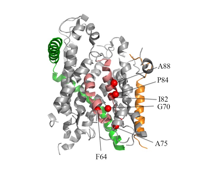
The bacterial Sec-dependent system is the major protein-biogenic pathway for protein secretion across the cytoplasmic membrane or insertion of integral membrane proteins into the phospholipid bilayer. The mechanism of SecA-driven protein transport across the SecYEG channel complex has remained controversial with conflicting claims from biochemical and structural studies regarding the depth and extent of SecA insertion into SecYEG during ongoing protein transport. Here we utilized site-specific in vivo photo-crosslinking to thoroughly map SecY regions that are in contact with SecA during its insertion cycle. An arabinose-inducible, rapidly folding OmpA-GFP chimera was utilized to jam the SecYEG channels with an arrested substrate protein to "freeze" them in their SecA-inserted state. Examination of 117 sites distributed throughout SecY indicated that SecA not only interacts extensively with the cytosolic regions of SecY as shown previously, but it also interacts with most of the transmembrane helices and periplasmic regions of SecY, with a clustering of interaction sights around the lateral gate and pore ring regions. Our observations support previous reports of SecA membrane insertion during in vitro protein transport as well as those documenting the membrane penetration properties of this protein. They suggest that one or more SecA regions transiently integrate into the heart of the SecY channel complex to span the membrane to promote the protein transport cycle. These findings indicate that high-resolution structural information about the membrane-inserted state of SecA is still lacking and will be critical for elucidating the bacterial protein transport mechanism.

Keywords: Sec system.


Conflict of interest statement

The authors declare that they have no conflicts of interest with the contents of this article

Figures

Figure 1.
Figure 1.
Experimental strategy. A, Sec61αγβ/SecYEG crystal structures from M. jannaschii (PDB code 1RHZ) (6) (upper row) or T. maritima (PDB 3DIN) (16) (lower row) highlighting the residues selected (depicted as colored spheres) for probing for SecA interaction within the SecY cytosolic loops as viewed from the top (i and iv), transmembrane helices as viewed from the side (ii and v), and periplasmic loops as viewed from the side (iii and vi). Sec61αγβ/SecYEG polypeptide chains are colored in gray, green, and orange, respectively. B, schematic diagram of SecA binding (left-hand side) and insertion (right-hand side) into the SecYEG channel during jamming caused by OmpA-GFP chimera production after arabinose induction.
Figure 2.
Figure 2.
In vivo photo-crosslinking analysis of SecY434 mutant during OmpA-GFP-induced translocon jamming. A, the E. coli Lys434 homologous residue is depicted as a hot pink sphere on the M. jannaschii Sec61αγβ/SecYEG crystal structure. Sec61αγβ/SecYEG polypeptide chains are colored in gray, green, and orange, respectively. B, titration of c-Myc-tagged SecY expression. The SecY434 mutant was grown in Miller broth supplemented with 1 mm pBpA and appropriate antibiotics up to an A600 of 0.15, when IPTG was added to the culture at the concentration indicated and cells were grown for another 60 min. Cells were harvested and membranes were isolated and analyzed by Western blotting with SecY (top) or c-Myc (bottom) peptide antibody as described under “Experimental procedures.” The positions of the c-Myc-tagged and chromosomally derived SecY protein are given. C-F, the SecY434 mutant was grown in Miller broth supplemented with 1 mm pBpA, 30 μm IPTG, 0.2% maltose and appropriate antibiotics to an A600 of 0.15, when 0.2% arabinose was added to a portion of the culture to induce OmpA-GFP jamming as indicated, and cells were harvested 45 min later. Cells were exposed to UV irradiation for 20 min as indicated, followed by cell breakage, membrane isolation, and analysis by Western blotting with: C, SecA antibody; D, GFP antibody; or E, c-Myc antibody as described under “Experimental procedures.” The positions of SecA and SecY proteins, the cross-linked SecA-SecY product (A-Y) or cross-linked SecY dimer (Y-Y), or OmpA-GFP chimera are given. Because of its rapidly folding GFP domain, OmpA-GFP usually runs as a doublet (39). F, Western blot analysis of OmpA-GFP-dependent translocon jamming kinetics of the SecY434 mutant. Cells were removed after arabinose addition at the indicated times and divided into cytoplasm–membrane (C) or periplasm (P) fractions and compared with the total cell (T) input by Western blotting using MBP antibody as described under “Experimental procedures.” The positions of pre-MBP and MBP are given. G, schematic diagram of the OmpA-GFP-jammed SecYEG channel showing positions of Cys21 at the end of the OmpA signal peptide and Cys68 within the plug domain of SecY that allows for efficient disulfide cross-linking of the SecY-jammed state. H, Western blot of the in vivo disulfide cross-linking experiment utilizing the Cys21 derivative of the OmpA-GFP chimera with a SecY mutant containing the Cys68 substitution. The strain was grown in Miller broth supplemented with 30 μm IPTG and appropriate antibiotics to A600 of 0.15, when 0.2% arabinose was added to a portion of the culture to induce OmpA-GFP jamming. Cells were harvested 45 min later and left untreated or treated with copper phenanthroline (CuPh3) in the absence or presence of DTT as described previously (40). The positions of SecY and the OmpA-GFP-SecY cross-linked product are indicated.
Figure 3.
Figure 3.
Mapping of SecA–SecY interaction sites by in vivo photo-crosslinking. Strains containing an amber mutation in the indicated SecY residue were grown with or without arabinose induction and subjected to UV irradiation as indicated followed by cell breakage, membrane isolation, and Western blotting with SecA antibody as described under “Experimental procedures.” Mutants were grouped according to the position of the amber mutation within each locale of SecY as follows: A, the cytosolic loop region; B, the transmembrane helix region; or C, the periplasmic loop region. The position of SecA and cross-linked SecA–SecY products (A-Y) are given along with stars that indicate the presumed partially degraded SecA–SecY products. Two representative mutants that were negative for SecA–SecY cross-linking are shown at the end of each panel. All SecY mutants were analyzed by photo-crosslinking at least three times with similar results in all cases.
Figure 3.
Figure 3.
Mapping of SecA–SecY interaction sites by in vivo photo-crosslinking. Strains containing an amber mutation in the indicated SecY residue were grown with or without arabinose induction and subjected to UV irradiation as indicated followed by cell breakage, membrane isolation, and Western blotting with SecA antibody as described under “Experimental procedures.” Mutants were grouped according to the position of the amber mutation within each locale of SecY as follows: A, the cytosolic loop region; B, the transmembrane helix region; or C, the periplasmic loop region. The position of SecA and cross-linked SecA–SecY products (A-Y) are given along with stars that indicate the presumed partially degraded SecA–SecY products. Two representative mutants that were negative for SecA–SecY cross-linking are shown at the end of each panel. All SecY mutants were analyzed by photo-crosslinking at least three times with similar results in all cases.
Figure 4.
Figure 4.
Location of constitutively positive SecA interaction sites within the lateral gate region of SecY. The M. jannaschii Sec61αγβ/SecYEG crystal structure is shown highlighting the E. coli SecY homologous residues Phe64, Gly70, Ala75, Ile82, Pro84, and Ala88 (red spheres) that were constitutively positive for cross-linking to SecA. The TM2b and TM7 helices within the lateral gate region are colored in pink, whereas the Sec61αγβ/SecYEG polypeptide chains are colored in gray, green, and orange, respectively.
Figure 5.
Figure 5.
Specificity of SecA antisera. A, the SecY434 mutant was grown with or without arabinose induction and subjected to UV irradiation as indicated followed by cell breakage, membrane isolation, and Western blotting with SecA antisera as described under “Experimental procedures.” For pre-adsorption of the antisera, 1 μl of SecA antisera was added to a 99-μl reaction containing 20 mm Tris-HCl, pH 7.5, 140 mm NaCl, 0.25% Tween 20 and the indicated amount of purified SecA protein, which was incubated on ice for 1 h prior to being used for Western blotting. The band highlighted by the diamond represents a nonspecific interaction given its behavior during immunoabsorption. B, comparison of the in vivo photo-crosslinking pattern of three SecY mutants by Western blotting utilizing SecA antisera or SecA antisera that was pre-adsorbed with 10 ng of purified SecA protein as indicated above.
Figure 6.
Figure 6.
Evaluation of cell lysis during the translocon jamming and in vivo photo-crosslinking procedure. The SecY58 mutant was grown with or without arabinose induction and subjected to UV irradiation as indicated under “Experimental procedures,” when samples were separated into supernatant and cell pellet fractions by sedimentation at 12,000 × g for 2 min at 4 °C. β-Galactosidase assays of the supernatant (S) and cell pellet (P) fractions were done in triplicate as described by Miller (55), and the standard deviation is shown.
Figure 7.
Figure 7.
Evaluation of the assembly state of SecY during in vivo photo-crosslinking. A, schematic diagram of a three-step sucrose density gradient indicating the floatation centrifugation positions of different membrane or protein species. B and C, the SecY391 mutant was grown with or without arabinose induction and subjected to UV irradiation as indicated followed by cell breakage and membrane isolation as described under “Experimental procedures.” B, samples were subjected to Western blotting with SecA antibody to assess the degree of SecA-SecY cross-linking. C, the isolated membrane fraction from the arabinose induced and UV-irradiated culture was adjusted to 1.74 m sucrose, and a 55-μl aliquot was loaded at the bottom of a centrifuge tube, and consecutively overlaid with 95 μl of 1.6 m sucrose followed by 75 μl of 1.25 m sucrose buffered with 50 mm Tris-HCl, pH 8, 50 mm KCl, 5 mm MgCl2. The sample was centrifuged at 98,000 rpm in a Sorvall S120AT3 rotor for 18 h at 4 °C. Eight equal fractions were collected from the top to bottom of the tube and analyzed by Western blotting using SecA (top), c-Myc (middle), or OmpA (bottom) antibody. The SecY391 mutant cells utilized for this experiment were the same as utilized for Fig. 3, and thus the image in B is the same as that of the corresponding image in Fig. 3B.
Figure 8.
Figure 8.
Jamming-dependent SecA-SecY cross-linking requires a functional signal peptide within the OmpA-GFP chimera. A, strains containing an amber mutation in the indicated SecY residue and an OmpA-GFP chimera with a functional (left-hand side) or non-functional (right-hand side) signal sequence were grown with arabinose induction and subjected to UV irradiation as indicated followed by cell breakage, membrane isolation, and Western blotting with SecA antibody as described under “Experimental procedures.” B, cells of the SecY58 mutant carrying the OmpA-RR-GFP chimera with a non-functional signal sequence were removed after arabinose addition at the indicated times and divided into cytoplasm–membrane (C) or periplasm (P) fractions and compared with the total cell (T) input by Western blotting using MBP antibody as described under “Experimental procedures.” C, comparison of the expression levels of the OmpA-GFP and OmpA-RR-GFP chimeras 45 min after arabinose induction for the SecY58 mutant by Western blotting using GFP antibody. The four mutant cultures utilized in this experiment were the same as utilized in Fig. 3, and thus the image in A is the same as the corresponding image in Fig. 3.
Figure 9.
Figure 9.
Structural summary of SecA-interactive SecY residues. SecY residues that scored positive for SecA interaction are depicted as spheres on the M. jannaschii (A) or T. maritima (B) Sec61αγβ/SecYEG crystal structure. Sec61αγβ/SecYEG polypeptide chains are colored gray, green, and orange, respectively. Positive residues are colored according to the degree of cross-linking found in our study with red, pink, and yellow indicating strong, medium, or weak cross-linking, respectively. Positive residues from the study of Mori and Ito (37) that are present in the corresponding two structures are colored purple. The dotted rectangle highlights the region around the lateral gate of SecY, which shows extensive interaction with SecA, whereas the dotted oval highlights the pore ring region, which showed numerous contacts with SecA during translocon jamming.

References

    1. du Plessis D. J., Nouwen N., and Driessen A. J. (2011) The Sec translocase. Biochim. Biophys. Acta 1808, 851–865 - PubMed
    1. Park E., and Rapoport T. A. (2012) Mechanisms of Sec61/SecY-mediated protein translocation across membranes. Annu. Rev. Biophys. 41, 21–40 - PubMed
    1. Ng D. T., Brown J. D., and Walter P. (1996) Signal sequences specify the targeting route to the endoplasmic reticulum membrane. J. Cell Biol. 134, 269–278 - PMC - PubMed
    1. Lee H. C., and Bernstein H. D. (2001) The targeting pathway of Escherichia coli presecretory and integral membrane proteins is specified by the hydrophobicity of the targeting signal. Proc. Natl. Acad. Sci. U.S.A. 98, 3471–3476 - PMC - PubMed
    1. Randall L. L., and Hardy S. J. (2002) SecB, one small chaperone in the complex milieu of the cell. Cell Mol. Life Sci. 59, 1617–1623 - PMC - PubMed

Publication types

MeSH terms

Substances

LinkOut - more resources