. 2017 Oct;10(5):e001706.
doi: 10.1161/CIRCGENETICS.117.001706.

Reduced Cardiac Calcineurin Expression Mimics Long-Term Hypoxia-Induced Heart Defects in Drosophila

Affiliations

Reduced Cardiac Calcineurin Expression Mimics Long-Term Hypoxia-Induced Heart Defects in Drosophila

Rachel Zarndt et al. Circ Cardiovasc Genet. 2017 Oct.

Erratum in

  • Correction.
    [No authors listed] [No authors listed] Circ Cardiovasc Genet. 2017 Dec;10(6):e000041. doi: 10.1161/HCG.0000000000000041. Circ Cardiovasc Genet. 2017. PMID: 30576608 No abstract available.

Abstract

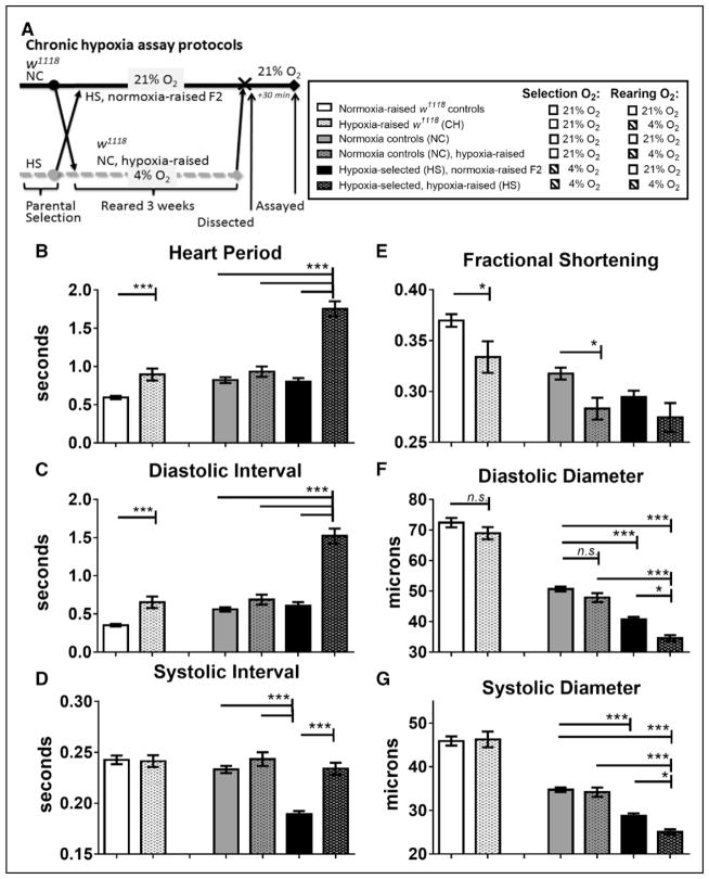
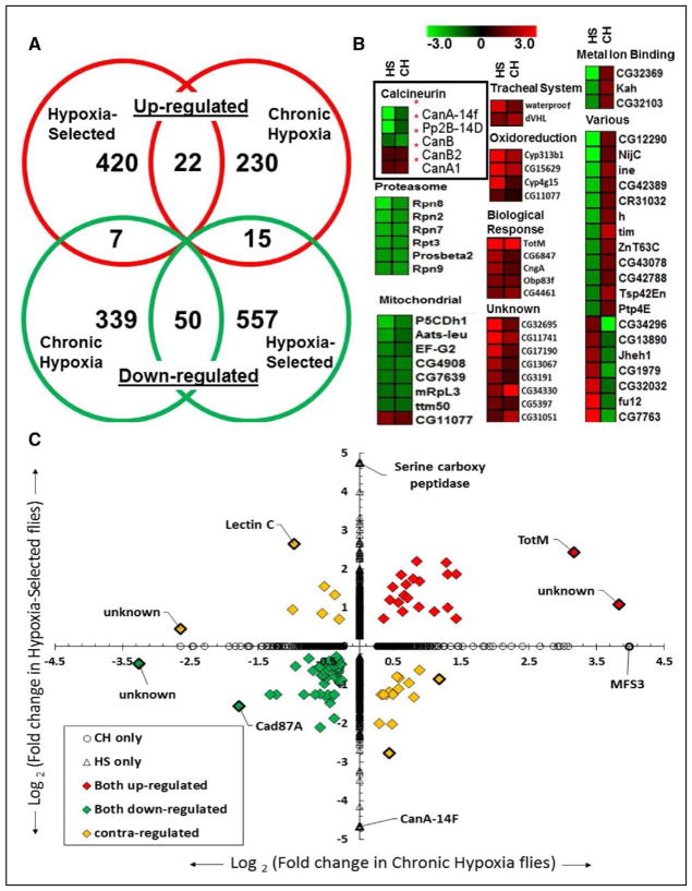
Background: Hypoxia is often associated with cardiopulmonary diseases, which represent some of the leading causes of mortality worldwide. Long-term hypoxia exposures, whether from disease or environmental condition, can cause cardiomyopathy and lead to heart failure. Indeed, hypoxia-induced heart failure is a hallmark feature of chronic mountain sickness in maladapted populations living at high altitude. In a previously established Drosophila heart model for long-term hypoxia exposure, we found that hypoxia caused heart dysfunction. Calcineurin is known to be critical in cardiac hypertrophy under normoxia, but its role in the heart under hypoxia is poorly understood.

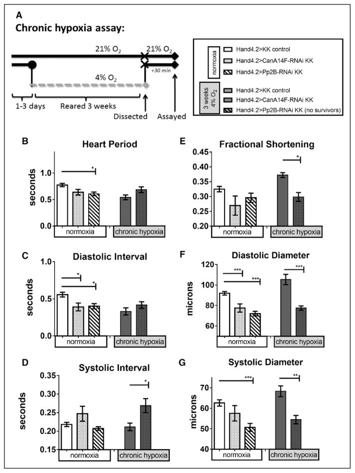
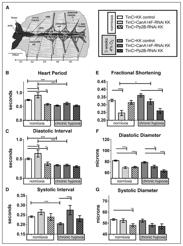
Methods and results: In the present study, we explore the function of calcineurin, a gene candidate we found downregulated in the Drosophila heart after lifetime and multigenerational hypoxia exposure. We examined the roles of 2 homologs of Calcineurin A, CanA14F, and Pp2B in the Drosophila cardiac response to long-term hypoxia. We found that knockdown of these calcineurin catalytic subunits caused cardiac restriction under normoxia that are further aggravated under hypoxia. Conversely, cardiac overexpression of Pp2B under hypoxia was lethal, suggesting that a hypertrophic signal in the presence of insufficient oxygen supply is deleterious.

Conclusions: Our results suggest a key role for calcineurin in cardiac remodeling during long-term hypoxia with implications for diseases of chronic hypoxia, and it likely contributes to mechanisms underlying these disease states.

Keywords: CanA; cardiac remodeling; cardiomyopathy; chronic ischemic heart disease; chronic mountain sickness; hypertrophy; hypoxia adaptation.


Figures

Figure 1
Figure 1
Cardiac response to chronic hypoxia in w1118 flies (CH) compared with hypoxia-selected flies (HS) and their normoxia control (NC) populations, reared under either hypoxia or normoxia. A, Protocol for chronic hypoxia treatment in female w1118 flies and in male HS and NC populations. B, Exposure of w1118 flies to chronic hypoxia exposure (CH) significantly increased heart period compared with their w1118 controls; HS flies reared under their chronic hypoxia selection conditions had heart periods significantly longer than either NC or HS population reared under normoxia or hypoxia-reared NC flies. C, Increase in heart period is caused by increases in diastolic intervals. D, Systolic intervals were unchanged after chronic hypoxia in all groups except normoxia-raised HS flies, where systolic interval is significantly decreased compared with NC and HS. E, Fractional shortening is significantly decreased in hearts from both w1118 flies and NC flies exposed to chronic hypoxia. F, Diastolic diameters were unchanged after chronic hypoxia in w1118 and NC, but were significantly reduced in normoxia-raised HS, compared with NC, and in hypoxia-raised HS compared with both NC and hypoxia-raised HS fly. G, Similarly, systolic diameters were unaffected by CH in w1118 or NC lines, but are reduced in normoxia-raised HS compared with NC and in hypoxia-raised HS compared with both NC and hypoxia-raised HS flies. All values are mean ± SEM for hearts (N=75, w1118 controls; N=41, hypoxia-raised w1118 (CH); N=57, NC; N=41, hypoxia-raised NC; N=60, normoxia-raised HS; N=36, HS). Two-way analysis of variance (ANOVA) with Tukey’s multiple comparisons post hoc test; n.s. indicates not significant, *P<0.05, ***P<0.001. Adapted from Zarndt et al with permission of the publisher. Copyright© 2015, The American Physiological Society. Authorization for this adaptation has been obtained both from the owner of the copyright in the original work and from the owner of copyright in the translation or adaptation.
Figure 2
Figure 2
Cardiac-specific differential gene expression in hypoxia-selected populations and in w1118 flies exposed to chronic hypoxia. Tran-scriptome analysis of RNA from isolated Drosophila hearts containing both myocardial and pericardial cells. A, Venn diagram depicting the number of genes differentially expressed in common or uniquely in hypoxia-selected flies (HS) compared with chronic hypoxia-exposed w1118 flies (CH) under 4% O2. B, Heat maps for groups of upregulated, downregulated, or contraregulated genes between HS and CH populations. The various categories include the 22 contraregulated genes between HS and CH populations. C, Log2-fold change in cardiac expression between HS vs CH flies with >1.2-fold upregulated (red), downregulated (green), or contraregulated genes and with P<0.05.
Figure 3
Figure 3
Effect of chronic hypoxia on heart function in hearts with myocardial and pericardial knockdown of calcineurins. A, left, Protocol for chronic hypoxia assay. BG, Heart function data under normoxia (left bars) or CH exposure (right bars) per genotypes indicated in figure key (A, right): progeny of CanA14F-RNAi and Pp2B-RNAi crossed to a Hand4.2-Gal4 driver causes knockdown (KD) specifically in pericardial and myocardial cells during development through adulthood. Hearts were dissected and assayed under 21% O2 after 3 weeks at 21% or 4% O2 (as in Zarndt et al). Note that cardiac Pp2B-RNAi KD flies under CH did not survive to 3 weeks. B, Heart period (HP) was reduced on cardiac Pp2B-RNAi KD in normoxia (condition: F=14.24, P=0.0002; genotype: F=8.089, P=0.0004; interaction=2.7, P=0.07). CanA14F KD had a similar put less pronounced effect. Under hypoxia, CanA14F KD had no significant effect. C, Similarly, diastolic intervals (DI) were reduced on cardiac CanA14F-RNAi and Pp2B-RNAi KD under normoxia, but not under CH (DI condition: F=13, P=0.0004; genotype: F=3.8, P=0.025; interaction =2.1, P=0.13). D, Systolic intervals (SI) were significantly elevated only in cardiac CanA14F KD flies after CH compared with controls under hypoxia (SI condition: F=0.64, P=0.43; genotype: F=7.8, P=0.0006; interaction =1.2, P=0.31). E, Fractional shortening (FS) was not significantly changed on cardiac calcineurin KD in normoxic flies. However, under hypoxia controls, CanA14F-RNAi KD in the heart results in reduced FS after CH (condition: F=0.53, P=0.47; genotype: F=20, P<0.0001; interaction=9.0, P=0.0002). F, Diastolic diameters were decreased significantly with both cardiac CanA14F-RNAi and PP2B-RNAi KD at normoxia, as well as with CanA14F KD under CH (condition: F=7.3, P=0.0009; genotype: F=14, P=0.0003; interaction=7.3, P=0.0009). G, Systolic diameters were similarly decreased with CanA14F and Pp2B KD at normoxia, as well as CanA14F KD under CH (condition: F=11, P=0.0009; genotype: F=1.4, P=0.26; interaction=F=3.0, P=0.055). All values are mean ± SEM (normoxia: N=37 KK control, N=33 CanA14F-RNAi, N=32 Pp2B-RNAi; chronic hypoxia: N=43 KK control, N=20 CanA14F-RNAi, N=20 Pp2B-RNAi). Two-way analysis of variance (ANOVA) and Sidak or Dunn multiple comparisons tests; *<0.05, **P<0.01, ***P<0.001. See also Materials and Methods.
Figure 4
Figure 4
Effect of chronic hypoxia on heart function in hearts with myocardial-specific knockdown of calcineurins. A, left, Diagram of Drosophila heart tube within dissected abdominal preparation (image courtesy of Dr. Georg Vogler). TinC-Gal4 drives expression specifically in myocardial cells (central tube), excluding the ostia (inflow valves). Hand4.2-Gal4-driven expression includes the ostia, as well as the pericardial cells. BG, Heart function data under normoxia (left bars) or CH exposure (right bars) per genotypes indicated in figure key (A, right): progeny of CanA14F-RNAi and Pp2B-RNAi crossed to a TinC-Gal4 driver causes knockdown (KD) specifically in myocardial cells during embryonic development and in pupal stages through adulthood. Hearts were dissected and assayed under 21% O2 after 3 weeks at 21% or 4% O2 (as in Zarndt et al). B, Heart period (HP) was elevated in CanA14F-RNAi but lowered in Pp2B-RNAi flies under normoxic conditions. After 3 weeks CH, HP was reduced for control and did not changed further on calcineurin KD (condition: F=14, P=0.0002; genotype: F=8.1, P=0.0004; interaction =2.7, P=0.070). C, Diastolic intervals changed similar to HP (condition: F=13, P=0.0004; genotype: F=3.8, P=0.025; interaction: F=2.1, P=0.13). D, Systolic intervals (SI) did not change on myocardial calcineurin KD under normoxia but were reduced in control flies after CH. Calcineurin KD in the myocardium under CH increased the systolic interval, similar to the Hand4.2-Gal4 data in Figure 3D (condition: F=0.64, P=0.43; genotype: F=7.8, P=0.0006; interaction: F=1.2, P=0.31). E, Fractional shortening (FS) was significantly reduced in myocardial CanA14F-RNAi KD, compared with controls, at normoxia and compared with CanA14F KD under CH. Myocardial Pp2B KD exhibited reduced FS on CH, but not under normoxia (condition: F=0.53, P=0.47; genotype: F=20, P<0.0001; interaction: F=9.0, P=0.0002). F, Diastolic diameters were decreased significantly with CanA14F-RNAi and Pp2B-RNAi KD under normoxia and CH (condition: F=7.3, P=0.0009; genotype: F=14, P=0.0003; interaction: F=7.3, P=0.0009). F, Systolic diameters were decreased, significantly only with Pp2B-RNAi KD at normoxia (condition: F=11, P=0.0009; genotype: F=1.4, P=0.26; interaction: F=3.0, P=0.055). All values are mean ± SEM (normoxia: N=32 KK control, N=30 CanA14F-RNAi, N=38 Pp2B-RNAi; chronic hypoxia: N=28 KK control, N=22 CanA14F-RNAi, N=20 Pp2B-RNAi). Data analysis: Kruskal–Wallis test and Dunn multiple comparisons post hoc test and 2-way analysis of variance (ANOVA) and Tukey’s multiple comparisons post hoc test; n.s.=not significant, *P<0.05, **P<0.01, ***P<0.001.

Comment in

References

    1. Prabhakar NR, Semenza GL. Adaptive and maladaptive cardiorespiratory responses to continuous and intermittent hypoxia mediated by hypoxia-inducible factors 1 and 2. Physiol Rev. 2012;92:967–1003. doi: 10.1152/physrev.00030.2011. - DOI - PMC - PubMed
    1. Monge C, León-Velarde F. Physiological adaptation to high altitude: oxygen transport in mammals and birds. Physiol Rev. 1991;71:1135–1172. - PubMed
    1. Ramirez JM, Folkow LP, Blix AS. Hypoxia tolerance in mammals and birds: from the wilderness to the clinic. Annu Rev Physiol. 2007;69:113–143. doi: 10.1146/annurev.physiol.69.031905.163111. - DOI - PubMed
    1. Penaloza D, Arias-Stella J. The heart and pulmonary circulation at high altitudes: healthy highlanders and chronic mountain sickness. Circulation. 2007;115:1132–1146. doi: 10.1161/CIRCULATIONAHA.106.624544. - DOI - PubMed
    1. Lankford HV, Swenson ER. Dilated hearts at high altitude: words from on high. High Alt Med Biol. 2014;15:511–519. doi: 10.1089/ham.2014.1047. - DOI - PubMed

Publication types

MeSH terms