. 2017 Oct 6;7(1):12775.
doi: 10.1038/s41598-017-13054-3.

Reactive metabolites of acetaminophen activate and sensitize the capsaicin receptor TRPV1

Affiliations

Reactive metabolites of acetaminophen activate and sensitize the capsaicin receptor TRPV1

Mirjam J Eberhardt et al. Sci Rep. .

Abstract

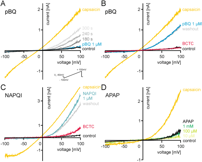
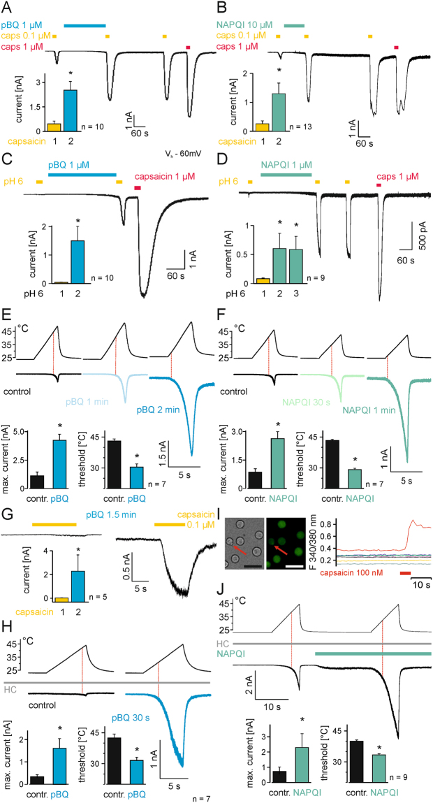
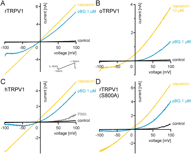
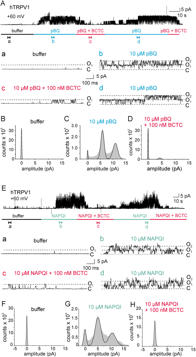
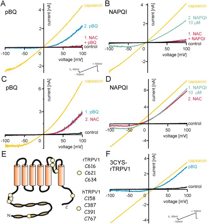
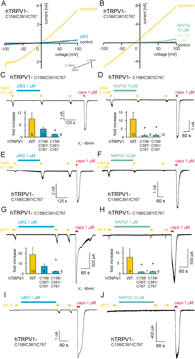
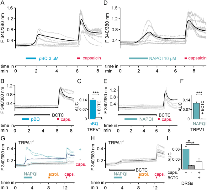
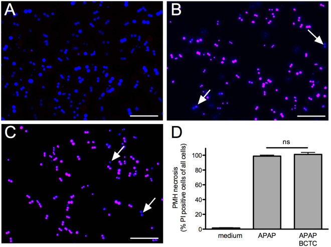
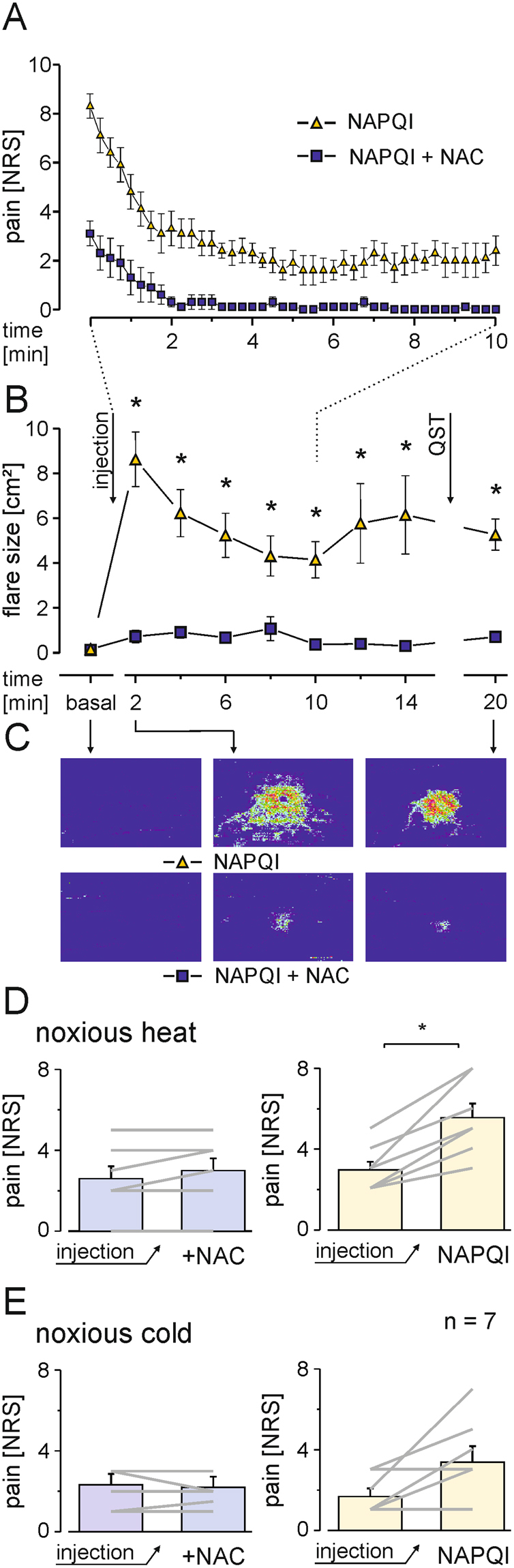
The irritant receptor TRPA1 was suggested to mediate analgesic, antipyretic but also pro-inflammatory effects of the non-opioid analgesic acetaminophen, presumably due to channel activation by the reactive metabolites parabenzoquinone (pBQ) and N-acetyl-parabenzoquinonimine (NAPQI). Here we explored the effects of these metabolites on the capsaicin receptor TRPV1, another redox-sensitive ion channel expressed in sensory neurons. Both pBQ and NAPQI, but not acetaminophen irreversibly activated and sensitized recombinant human and rodent TRPV1 channels expressed in HEK 293 cells. The reducing agents dithiothreitol and N-acetylcysteine abolished these effects when co-applied with the metabolites, and both pBQ and NAPQI failed to gate TRPV1 following substitution of the intracellular cysteines 158, 391 and 767. NAPQI evoked a TRPV1-dependent increase in intracellular calcium and a potentiation of heat-evoked currents in mouse spinal sensory neurons. Although TRPV1 is expressed in mouse hepatocytes, inhibition of TRPV1 did not alleviate acetaminophen-induced hepatotoxicity. Finally, intracutaneously applied NAPQI evoked burning pain and neurogenic inflammation in human volunteers. Our data demonstrate that pBQ and NAQPI activate and sensitize TRPV1 by interacting with intracellular cysteines. While TRPV1 does not seem to mediate acetaminophen-induced hepatotoxicity, our data identify TRPV1 as a target of acetaminophen with a potential relevance for acetaminophen-induced analgesia, antipyresia and inflammation.


Conflict of interest statement

The authors declare that they have no competing interests.

Figures

Figure 1
Figure 1
Ramp currents generated by TRPV1 are sensitized by pBQ and NAPQI. Traces show voltage ramp-induced membrane currents (see insert) in hTRPV1-expressing HEK 293 cells, one representative recording out of 6–7 measured cells of each group. (A) Sensitization of hTRPV1 by 1 µM pBQ is dependent on the duration of application, a maximum is reached at about 300 s. 1 µM capsaicin was used as control. (B) 100 nM BCTC blocks pBQ (5 min)-induced currents. Note that currents slowly recover following washout of BCTC for 2 minutes. (C) 1 µM NAPQI (5 min) sensitizes ramp currents generated by TRPV1, and these can be transiently blocked by BCTC. (D) Increasing concentrations of acetaminophen (APAP 10 µM, 100 µM, 1000 µM, each applied for 4 minutes) has no effects on ramp currents evoked in hTRPV1-expressing cells.
Figure 2
Figure 2
pBQ and NAPQI sensitize TRPV1.Inward currents evoked by 100 nM capsaicin are constantly increased after application of 1 µM pBQ (A) or 10 µM NAPQI (B) for about 240 s even without their further presence. Proton-induced responses are also sensitized by application of pBQ (C) and NAPQI (D). Bar diagrams show means ( ± SEM) of capsaicin- (A,B) or proton-induced (C,D) inward currents (*p ≤ 0.02). pBQ (E) and NAPQI (F) shift the temperature threshold of currents induced by heat ramps (25–45 °C) to lower temperatures, shown as red dotted lines in representative recordings and as bar diagrams of temperature needed to evoke inward currents before and after treatment with pBQ (E) and NAPQI (F). The maximum heat-induced inward currents are also increased by both metabolites (pBQ *p = 0.028; NAPQI *p = 0.018). Similarly, in DRG neurons of mice pBQ sensitized capsaicin (100 nM) -induced (G) and heat induced-currents (H) and shifted temperature thresholds to lower temperatures. 50 µM HC 030031 was used throughout these experiments to block TRPA1 (*p = 0.043 (G); *p = 0.018 (H)). (I) A 100 nM capsaicin stimulus was used to identify DRG neurons expressing TRPV1. From left to right: bright field image of small DRG neurons; same neurons loaded with FURA-2AM excited at 340 nm (scale bar = 50 µm); traces of cells during a short application of 100 nM capsaicin. The capsaicin-responding neuron is marked in red and labeled with an arrow. (J) Heat-induced currents in TRPV1-expressing neurons are immediately sensitized by 10 µM NAPQI, note that NAPQI itself evokes a small inward current, while TRPA1 was blocked by HC 030031. NAPQI increased heat induced inward currents and shifted temperature thresholds of TRPV1 to lower temperatures (*p = 0.018).
Figure 3
Figure 3
Activation of TRPV1 by pBQ does not involve the capsaicin-binding domain or proteinkinase C-dependent phosphorylation. pBQ (1 µM, 1.5–2 min) sensitized voltage ramp-induced currents in rTRPV1 (A) and oTRPV1 (B), the latter suggesting a mechanism independent of the capsaicin-binding site. (C) Sensitization of hTRPV1 by pBQ (1 µM, 2 min) is additive to sensitization by PKC-dependent phosphorylation evoked by PMA (300 nM). (D) In an rTRPV1 mutant lacking the PKC-phosphorylation site S800, membrane currents are still clearly sensitized by pBQ (1 µM 2–3 min).
Figure 4
Figure 4
pBQ and NAPQI directly activate TRPV1 from the intracellular side. (A) Single channel recordings from inside-out patches show an increased open state probability of TRPV1 channels during 10 µM pBQ application (b) which is decreased by co-application of 100 nM BCTC (c). Sections a,b,c and d represent episodes of 1 s of the upper trace with an expanded time scale. (B,C,D) Amplitude histograms calculated from 20 s sections of the full trace in Fig. 4A show two peaks for the open states of two channels in the patch and a small closed state peak during application of pBQ (C). Histograms (B and D) display a prominent closed state probability of TRPV1 channels before pBQ treatment (buffer, B) and after pBQ treatment with co-application of BCTC (D). (E) Representative trace for single channel recordings from inside-out patches with increased open state probability of TRPV1 during application of 10 µM NAPQI (b) and decreased open state probability by co-application of 100 nM BCTC (c) Sections a, (b,c) and d represent 1 s episodes of the upper trace with an expanded time scale. (F,G) and (H). Amplitude histograms calculated from 20 s sections of the full trace in Fig. 4E showing two peaks for the open states of two channels in the patch and a small closed state peak during application of NAPQI (G). TRPV1 channels reside predominantly in the closed state before NAPQI treatment (buffer, F) and during co-application of NAPQI and BCTC (H).
Figure 5
Figure 5
Modulation of cysteines underlies the sensitization of TRPV1 by pBQ and NAPQI. Co-application of 1 mM NAC (5 min) prevents sensitization of ramp currents in hTRPV1 by pBQ (A) and NAPQI (B). Note that a second application of pBQ (1 µM, 3 min) or NAPQI (10 µM, 1.5 min) alone again increases ramp currents. When NAC (1 mM, 4 min) is applied after sensitization of currents has already been evoked, it fails to reverse the effects of pBQ (C) and NAPQI (D). (E) Schematic model of TRPV1: external cysteines important for sensitization of rTRPV1 by reducing and oxidizing agents close to the pore forming region are marked with green dots (3CYS-rTRPV1). Internal cysteines of hTRPV1 which have been described to be modulated by oxidative challenges leading to channel sensitization are marked in yellow. (F) pBQ strongly sensitizes the rTRPV1 mutant lacking external cysteines.
Figure 6
Figure 6
Modulation of cysteines underlies the sensitization of TRPV1 by pBQ and NAPQI. (A,B) Mutation of internal cysteines in hTRPV1 abolishes sensitization of by both pBQ (G C158A/C391S/C767S-hTRPV1) and NAPQI (H C158A/C387S/C767S-hTRPV1). pBQ and both concentrations of NAPQI were applied for five minutes. These cysteines are also involved in sensitization of capsaicin- (CF) and proton-induced (GJ) inward currents by both metabolites. Representative traces are displayed for pBQ (C and G, C158S/C391S/C767S-hTRPV1; (E and I) C158A/C387S/C767S-hTRPV1) and NAPQI (D and H), C158A/C387S/C767S-hTRPV1; F and J, C158S/C391S/C767S-hTRPV1). Bar diagrams show multiples of capsaicin- or proton-induced responses before and after application of pBQ (C and G) or NAPQI (D and H) in wild-type and mutant hTRPV1. Sensitization of capsaicin- and proton-induced responses was reduced for pBQ in C158A/C391S/C767S-hTRPV1 (*p ≤ 0.007), and completely lost for NAPQI in both mutants (*p ≤ 0.0009 respectively *p ≤ 0.005).
Figure 7
Figure 7
pBQ and NAPQI induce a TRPV1-dependent rise in intracellular calcium. (A) In hTRPV1-expressing HEK 293 cells, 3 µM pBQ induced a rise in intracellular calcium in 36% of cells which also responded to capsaicin (n = 392). (B) Increase in intracellular calcium by pBQ was inhibited by the TRPV1 blocker BCTC (100 nM; n = 340). Responses are shown as mean of all cells responsive to pBQ (A) or capsaicin (B bold black trace), and calcium measurements of representative cells (thin grey traces). (C) Areas under the curve (AUC) of evoked calcium signals of all cells responding to pBQ: pBQ responses (blue bar) were significantly reduced by the TRPV1 channel blocker BCTC (white bar p < 0.001). (D) Similar to pBQ, 10 µM NAPQI evoked increases in intracellular calcium in hTRPV1-expressing cells (59%, n = 282). (A) These responses were inhibited by BCTC in 95% of capsaicin-responsive cells (n = 602). Bold black trace (mean) and thin grey traces (representative cells) of calcium measurements. (F) AUC of evoked calcium signals of all cells measured responding to NAPQI: NAPQI responses (cyan bar) were significantly reduced by BCTC (white bar; p < 0.001). (G) 10 µM NAPQI induced an increase in intracellular calcium in 15% of DRG neurons from TRPA1-knockout mice. Calcium responses of capsaicin-responding (blue, n = 216) and non-responding (gray, n = 166) subgroups of DRG neurons (bold = mean, thin = representative measurements of each group). (H) Co-application of the TRPV1 blocker BCTC reduced NAPQI-evoked calcium responses in DRG neurons from TRPA1-knockout mice (bold = mean of capsaicin-sensitive neurons, thin grey traces display representative measurements). (I) Comparison of the AUCs of calcium increase of the traces presented in (G and H). Increases in intracellular calcium following application of NAPQI are significantly inhibited in cells unresponsive to capsaicin (gray bar) or by TRPV1 block by BCTC (white bar; p = 0.00002).
Figure 8
Figure 8
Block of TRPV1 does not inhibit acetaminophen-induced hepatotoxicity. (AC) Primary mouse hepatocytes were treated with medium (A), 10 mM acetaminophen (B) or acetaminophen and 10 µM BCTC (C) for 22 h and stained with propidium iodide (PI, red) to visualize necrotic cells and DAPI (blue) for staining the nucleus of all cells. In (A) there are only blue colored nuclei visible indicating the absence of necrotic cells. Whereas the treatment with acetaminophen (B) even in combination with BCTC (C) resulted in almost 100% necrotic cells that are DAPI and PI positive and appeared as purple colored nuclei (arrows point to DAPI positive and PI negative cell nuclei; scale bar = 100 µM). (D) Bar graphs indicate percentage of necrotic cells following treatment of mouse hepatocytes with 10 mM acetaminophen, the TRPV1 blocker BCTC at 10 µM is not efficient to reduce or prevent cell death.
Figure 9
Figure 9
Injection of NAPQI in human skin induces pain and axon-reflex erythema. (A) Magnitude and time course of NAPQI-evoked pain in human volunteers after intracutaneous injection to the volar forearm. Pain was rated on a numerical rating scale (NRS) from 0 to 10. With co-application NAC (10 mM), pain following injection of NAPQI was reduced (n = 7, mean ± SEM). (B) Laser Doppler scanning to measure superficial blood flow reveals increased flow after NAPQI injection in comparison to NAPQI and NAC (p ≤ 0.011). (C) Representative pseudocolor image series of NAPQI ± NAC injections in one volunteer. (D) Pain ratings to noxious heat (47 °C, 10 s) increase following intracutaneous injection of NAPQI, but not when co-applied with NAC. Diagrams show responses before after injection of NAPQI alone (right) or in combination with NAC (left) presented as mean ± SEM and responses of all subjects tested (grey lines; *p = 0.018). (E) Only in some volunteers NAPQI alone (right), but not in combination with NAC (left) also increased responses to noxious cold (0 °C, 10 s; p = 0.069).

References

    1. Hinz B, Cheremina O, Brune K. Acetaminophen (paracetamol) is a selective cyclooxygenase-2 inhibitor in man. FASEB journal: official publication of the Federation of American Societies for Experimental Biology. 2008;22:383–390. doi: 10.1096/fj.07-8506com. - DOI - PubMed
    1. Mitchell JR, Jollow DJ, Potter WZ, Gillette JR, Brodie BB. Acetaminophen-induced hepatic necrosis. IV. Protective role of glutathione. The Journal of pharmacology and experimental therapeutics. 1973;187:211–217. - PubMed
    1. Kerckhove N, et al. Ca(v)3.2 calcium channels: the key protagonist in the supraspinal effect of paracetamol. Pain. 2014;155:764–772. doi: 10.1016/j.pain.2014.01.015. - DOI - PubMed
    1. Pickering G, Esteve V, Loriot MA, Eschalier A, Dubray C. Acetaminophen reinforces descending inhibitory pain pathways. Clinical pharmacology and therapeutics. 2008;84:47–51. doi: 10.1038/sj.clpt.6100403. - DOI - PubMed
    1. Andersson DA, et al. TRPA1 mediates spinal antinociception induced by acetaminophen and the cannabinoid Delta(9)-tetrahydrocannabiorcol. Nature communications. 2011;2:551. doi: 10.1038/ncomms1559. - DOI - PubMed

Publication types

MeSH terms

LinkOut - more resources