. 2017:1677:1-80.
doi: 10.1007/978-1-4939-7321-7_1.

NMDA Receptors in the Central Nervous System

Affiliations

NMDA Receptors in the Central Nervous System

Kasper B Hansen et al. Methods Mol Biol. 2017.

Abstract

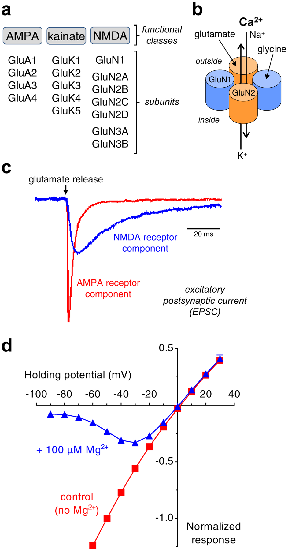
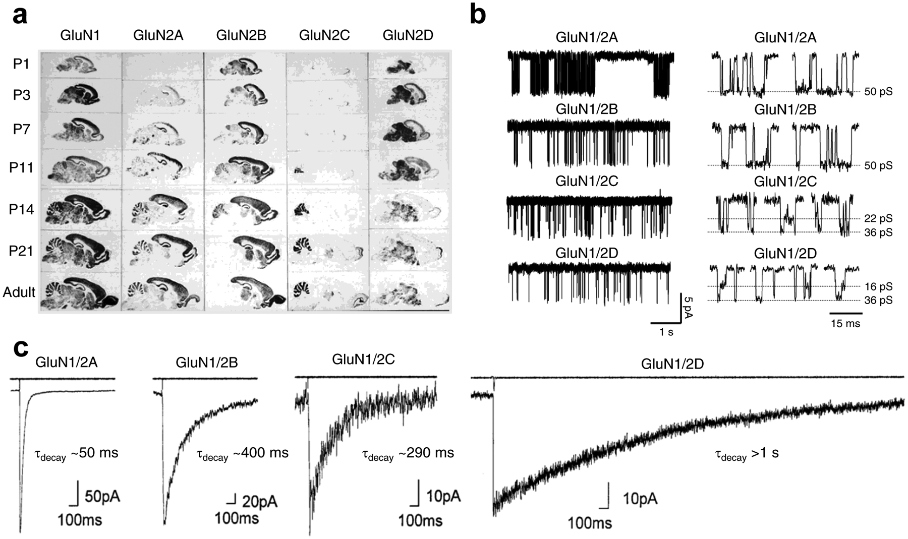
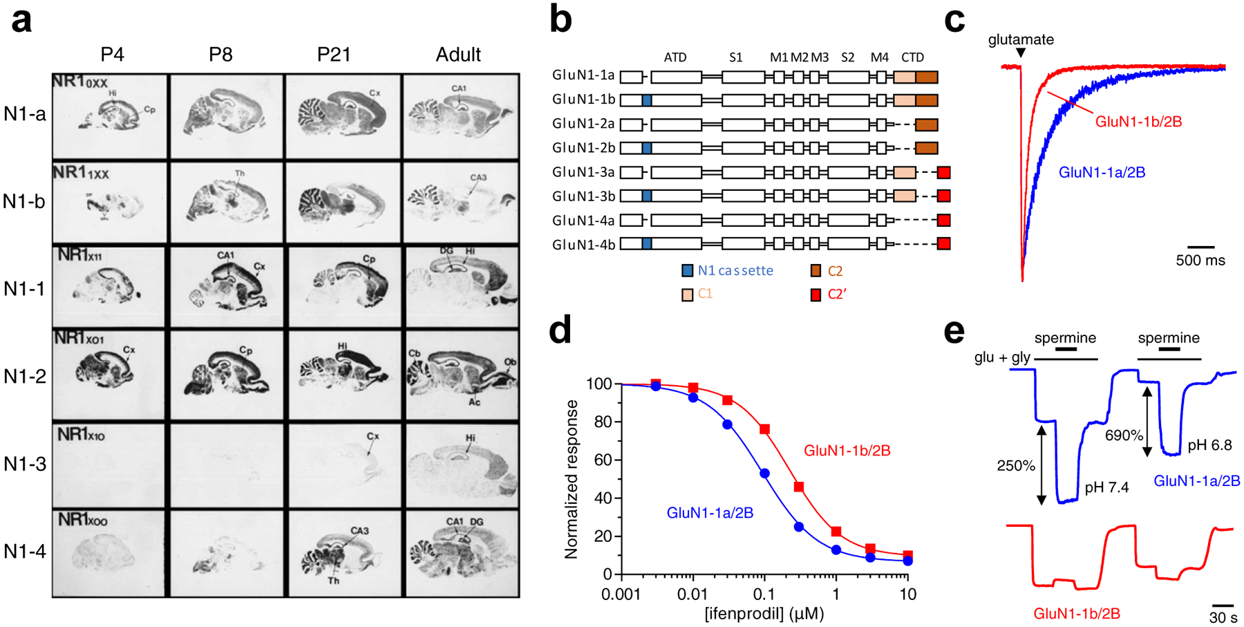
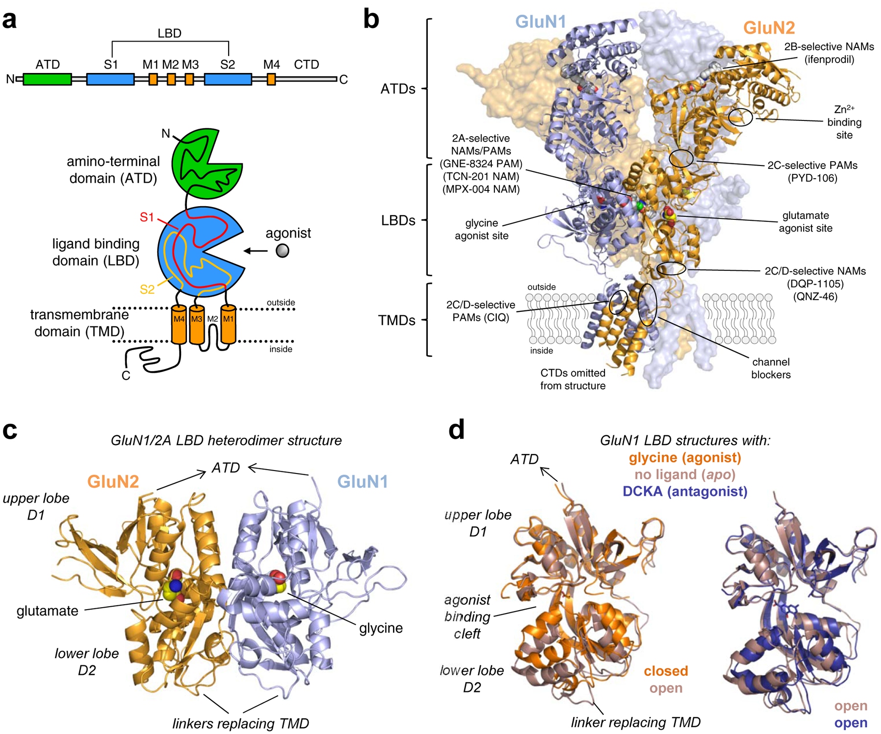
NMDA-type glutamate receptors are ligand-gated ion channels that mediate a major component of excitatory neurotransmission in the central nervous system (CNS). They are widely distributed at all stages of development and are critically involved in normal brain functions, including neuronal development and synaptic plasticity. NMDA receptors are also implicated in the pathophysiology of numerous neurological and psychiatric disorders, such as ischemic stroke, traumatic brain injury, Alzheimer's disease, epilepsy, mood disorders, and schizophrenia. For these reasons, NMDA receptors have been intensively studied in the past several decades to elucidate their physiological roles and to advance them as therapeutic targets. Seven NMDA receptor subunits exist that assemble into a diverse array of tetrameric receptor complexes, which are differently regulated, have distinct regional and developmental expression, and possess a wide range of functional and pharmacological properties. The diversity in subunit composition creates NMDA receptor subtypes with distinct physiological roles across neuronal cell types and brain regions, and enables precise tuning of synaptic transmission. Here, we will review the relationship between NMDA receptor structure and function, the diversity and significance of NMDA receptor subtypes in the CNS, as well as principles and rules by which NMDA receptors operate in the CNS under normal and pathological conditions.

Keywords: Disease; Ion channel; Ionotropic glutamate receptor; NMDA; Neurotransmitter; Regulation; Structure-function; Synaptic transmission.


Figures

Figure 1.
Figure 1.. Functional classes of ionotropic glutamate receptors.
a) Ionotropic glutamate receptors are divided into three functional classes, namely AMPA, kainate, and NMDA receptors. Multiple subunits have been cloned in each of these classes. b) The majority of NMDA receptors in the CNS are composed of two glycine-binding GluN1 and two glutamate-binding GluN2 subunits, which form a central cation-permeable channel pore. c) AMPA and NMDA receptor-mediated components of the EPSC at a central synapse. The slow NMDA receptor-mediated component is isolated in the absence of Mg2+ using the AMPA receptor antagonist CNQX, whereas the fast AMPA receptor-mediated component is isolated using the NMDA receptor antagonist AP5. The figure shows unpublished data from Lonnie P. Wollmuth and is adapted with permission from Traynelis et al. [1]. d) Relationship between NMDA receptor current response and membrane potential (i.e. I/V-relationship) in the presence and absence of 100 μM extracellular Mg2+. Voltage-dependent Mg2+-block is relieved with depolarization of the membrane potential (i.e. as the membrane potential approaches 0 mV). Unpublished data from Feng Yi and Kasper B. Hansen.
Figure 2.
Figure 2.. GluN2 subunit-specific expression and functional properties of recombinant NMDA receptor subtypes.
a) Regional and developmental expression of GluN2 subunits in rat brain revealed in autoradiograms using in situ hybridizations of oligonucleotide probes for the relevant mRNAs to parasagittal sections. Modified with permission from Akazawa et al. [92]. b) Single-channel recordings of currents from diheteromeric NMDA receptor subtypes expressed in HEK293 cells (outside-out membrane patches). Open probability is ~0.5 for GluN1/2A, ~0.1 for GluN1/2B, and <0.05 for GluN1/2C and GluN1/2D. Highlights of individual openings are shown on the left. GluN1/2A and GluN1/2B have higher channel conductance (~50 pS) compared to GluN1/2C (~22 and ~36 pS) and GluN1/2D (~16 and ~36 pS). Adapted with permission from Yuan et al. [524]. c) Whole-cell patch-clamp recordings of responses from brief application of glutamate (1 ms of 1 mM glutamate) to recombinant diheteromeric NMDA receptor subtypes expressed in HEK293 cells. The open tip current indicating the duration of the drug application is shown in the upper trace. Adapted with permission from Vicini et al. [62].
Figure 3.
Figure 3.. Expression and functional properties of GluN1 splice variants.
a) Regional and developmental expression of GluN1 splice variants in rat brain revealed in autoradiograms using in situ hybridizations of oligonucleotide probes for the relevant mRNAs to parasagittal sections. Ac, nucleus accumbens; Cb, cerebellum; Cp, caudate-putamen; Cx, cortex; DG, dentate gyrus; DP, dorsal pons; Hi, hippocampus; Ob, olfactory bulb; Th, thalamus; VPn, ventro-posterial thalamic nuclei. Modified with permission from Paupard et al. [78]. b) Linear representation of the GluN1 polypeptide chain for eight alternative splice variants. GluN1 subunits are composed of the amino-terminal domain (ATD), S1 and S2 segments that form the ligand binding domain (LBD), three transmembrane helices (M1, M3, and M4) and a membrane reentrant loop (M2), and the intracellular carboxyl-terminal domain (CTD). The N1 cassette (blue) is 21 amino acids in the ATD encoded by exon 5. The C1 cassette (yellow) is 37 amino acids in the CTD encoded by exon 21, while the C2 cassette (orange) is 38 amino acids in the CTD encoded by exon 22. Deletion of exon 22 creates a shift in the open reading frame, resulting in the alternate exon 22’ that encodes the C2’ cassette (red; 22 amino acids). c) Whole-cell patch-clamp recordings of responses from brief application of glutamate (1 ms of 1 mM glutamate) to recombinant GluN1–1a/2B and GluN1–1b/2B receptors expressed in HEK293 cells. NMDA receptors containing exon 5 (e.g. as in GluN1–1b) display faster deactivation time course compared to receptors lacking exon 5 (e.g. as in GluN1–1a). d) Ifenprodil concentration-inhibition relationships for recombinant GluN1–1a/2B and GluN1–1b/2B receptors expressed in Xenopus oocytes. Ifenprodil potency is lower for receptors containing exon 5. e) Representative recordings for spermine potentiation of responses from recombinant GluN1–1a/2B and GluN1–1b/2B receptors expressed in Xenopus oocytes. Spermine sensitivity is dramatically reduced for receptors containing exon 5. Data in c-e) are unpublished from Feng Yi and Kasper B. Hansen.
Figure 4.
Figure 4.. Functional properties of triheteromeric GluN1/2A/2B receptors.
a) Ifenprodil concentration-inhibition relationships for recombinant diheteromeric GluN1/2A and GluN1/2B receptors and triheteromeric GluN1/2A/2B receptors expressed in Xenopus oocytes using a method to control subunit composition of NMDA receptors [151]. Ifenprodil efficacy and potency are reduced for triheteromeric GluN1/2A/2B receptors that only contain one binding site for ifenprodil. b) Whole-cell patch-clamp recordings of responses from brief application of glutamate (1 ms of 1 mM glutamate) to recombinant diheteromeric GluN1/2A and GluN1/2B receptors and triheteromeric GluN1/2A/2B receptors expressed in HEK293 cells. The deactivation time course of triheteromeric GluN1/2A/2B receptors is similar to diheteromeric GluN1/2A and strikingly different from diheteromeric GluN1/2B. Data are adapted with permission from Hansen et al. [151].
Figure 5.
Figure 5.. NMDA receptor structure and ligand binding sites.
a) Linear representation and cartoon illustration of the polypeptide chain in iGluR subunits. Each subunit consists of a large extracellular amino-terminal domain (ATD), a bi-lobed ligand binding domain (LBD), a transmembrane domain (TMD), and an intracellular CTD. The TMD is formed by three transmembrane helices (M1, M2, and M4) and a membrane re-entrant loop (M2). The LBD is formed by two segments of the polypeptide chain (S1 and S2), which fold into a kidney-shaped structure composed of an upper lobe (D1) and lower lobe (D2) relative to the cell membrane, and the agonist binding site is located in the cleft between the two lobes. b) Crystal structure of the GluN1/2B NMDA receptor (PDB ID 4PE5; [67]), illustrating the subunit arrangement and the layered domain organization composed of the TMD layer and two extracellular layers formed by LBDs and ATDs. Agonist binding sites as well as known and predicted binding sites for positive and negative allosteric modulators (PAMs and NAMs) are highlighted. c) Crystal structure of the soluble GluN1/2A LBD heterodimer (PDB ID 5I57; [158]), showing the subunit interface and back-to-back dimer arrangement of the LBDs. Soluble LBD proteins composed of the S1 and S2 segments of the polypeptide chain are produced by deleting the ATD and replacing the TMD with a di-peptide linker. d) Overlay of crystal structures of the soluble GluN1 LBD in the apo-form (PDB 4KCC; [168]) or in complex with the agonist glycine (PDB ID 5I57; [158]) or competitive antagonist DCKA (PDB ID 4NF4; [166]). The upper D1 lobes are aligned to illustrate the similar conformations of antagonist-bound and apo-form structures. Agonist binding induces considerable closure of the LBD compared to the antagonist-bound and apo-form structures, and agonist-induced closure of the LBD is required for activation of NMDA receptors. Competitive antagonists bind the LBD without inducing domain closure, thereby preventing agonist binding and receptor activation.
Figure 6.
Figure 6.. Subunit crossover and symmetry mismatch in the NMDA receptor structure.
Side view of the GluN1/2B NMDA receptor structure (PDB ID 4PE5; [67]) and top views of the ATD, LBD, and TMD layers. The subunits in GluN1/2 receptors are arranged in an alternating pattern (i.e. 1-2-1-2) and there is a symmetry mismatch between the TMDs and the extracellular LBDs and ATDs of the receptor. The TMDs are arranged symmetrically around the ion channel pore with a quasi-4-fold symmetry, whereas the extracellular portion adopts a dimer-of-dimer arrangement (i.e. two GluN1/2 heterodimers) with a 2-fold symmetry. There is a subunit crossover between the LBD layer and the ATD layer in that the GluN1(α) ATD forms a local dimer with the GluN2B(α) ATD, whereas the GluN1(α) LBD forms a local dimer with the GluN2B(β) LBD.
Figure 7.
Figure 7.. Structural determinants in the NMDA receptor ion channel pore.
a) View parallel to the membrane of the TMDs in the GluN1/2B NMDA receptor structure (PDB ID 4TLM; [66]). The solvent accessible surface is carved along the pore axis using the computer program HOLE and shows the M3 bundle crossing near the extracellular side of the membrane, which presumably forms the activation gate, and the narrow constriction in the pore (Q/R/N site). Green dots indicate a pore radius of 1.15–2.3 Å and blue dots indicate a pore radius greater than 2.3 Å. b) View of the TMDs from the extracellular side of the membrane along the pore axis. GluN1 and GluN2B subunits are blue and orange, respectively. The α-carbon of residues T646 and A645, which appear to define the activation gate, are highlighted as spheres. Adapted with permission from Lee et al. [66].

References

    1. Traynelis SF, Wollmuth LP, McBain CJ, Menniti FS, Vance KM, Ogden KK, Hansen KB, Yuan H, Myers SJ, Dingledine R (2010) Glutamate receptor ion channels: structure, regulation, and function. Pharmacol Rev 62 (3):405–496. doi: 10.1124/pr.109.002451 - DOI - PMC - PubMed
    1. Paoletti P, Bellone C, Zhou Q (2013) NMDA receptor subunit diversity: impact on receptor properties, synaptic plasticity and disease. Nat Rev Neurosci 14 (6):383–400. doi: 10.1038/nrn3504 - DOI - PubMed
    1. Matsuda K, Yuzaki M (2012) Cbln1 and the delta2 glutamate receptor--an orphan ligand and an orphan receptor find their partners. Cerebellum 11 (1):78–84. doi: 10.1007/s12311-010-0186-5 - DOI - PubMed
    1. Yuzaki M (2009) New (but old) molecules regulating synapse integrity and plasticity: Cbln1 and the delta2 glutamate receptor. Neuroscience 162 (3):633–643 - PubMed
    1. Schmid SM, Hollmann M (2008) To Gate or not to Gate: Are the Delta Subunits in the Glutamate Receptor Family Functional Ion Channels? Mol Neurobiol 37 (2–3):126–141 - PubMed

Publication types

MeSH terms

Substances