. 2017:1677:145-162.
doi: 10.1007/978-1-4939-7321-7_7.

Selective Cell-Surface Expression of Triheteromeric NMDA Receptors

Affiliations

Selective Cell-Surface Expression of Triheteromeric NMDA Receptors

Feng Yi et al. Methods Mol Biol. 2017.

Abstract

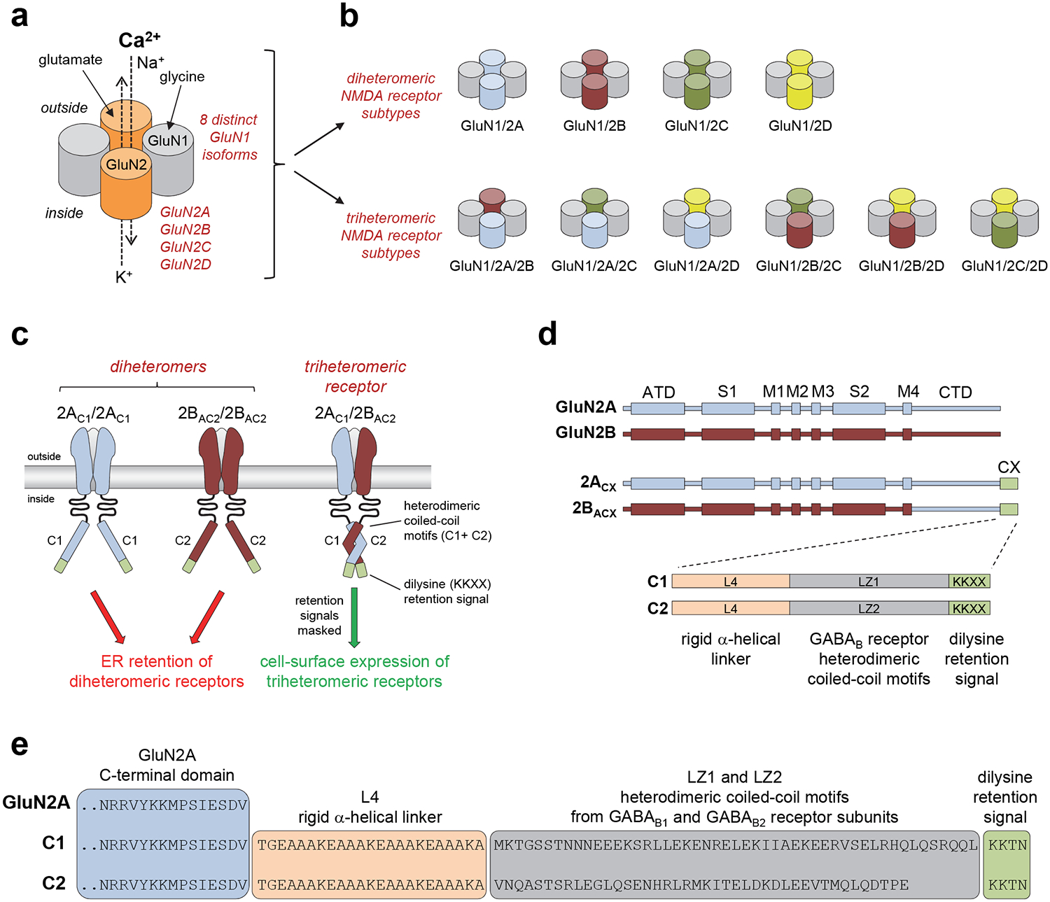
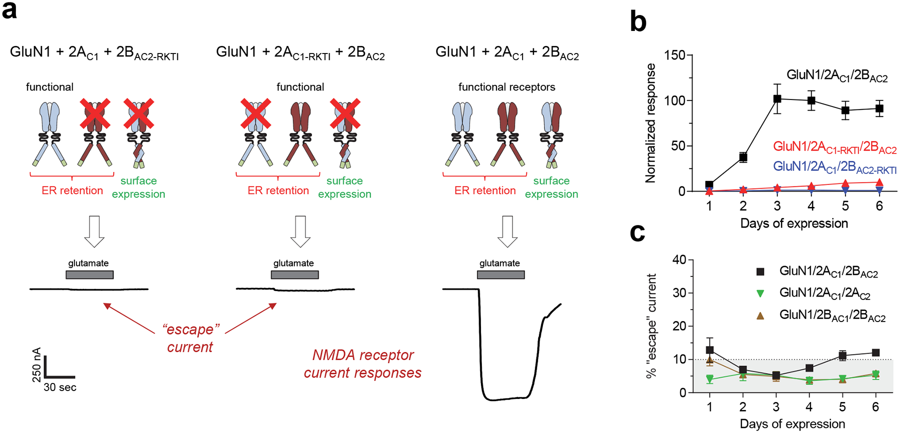
The NMDA-type ionotropic glutamate receptors play pivotal roles in many brain functions, but are also involved in numerous brain disorders. Seven NMDA receptor subunits exist (GluN1, GluN2A-D, and GluN3A-B) that assemble into a diverse array of tetrameric receptor subtypes with distinct functional properties and physiological roles. Most NMDA receptors are composed of two GluN1 and two GluN2 subunits, which can assemble into four diheteromeric receptor subtypes composed of GluN1 and one type of GluN2 subunit (e.g., GluN1/2A), and presumably also six triheteromeric receptor subtypes composed of GluN1 and two different GluN2 subunits (e.g., GluN1/2A/2B). Despite accumulating evidence that a large proportion of native NMDA receptors are triheteromers, little is known about their function and pharmacology due to the lack of methods to faithfully express triheteromeric NMDA receptors in heterologous expression systems. The problem is that co-expression of GluN1 with two different GluN2 subunits generates two distinct diheteromeric receptor subtypes as well as one triheteromeric receptor subtype, thereby confounding studies on a homogenous population of triheteromeric NMDA receptors. Here, we will describe a method to selectively express recombinant triheteromeric GluN1/2A/2B receptors without interfering co-expression of diheteromeric GluN1/2A and GluN1/2B receptors. This method enables quantitative evaluation of functional and pharmacological properties of triheteromeric GluN1/2A/2B receptors, which are presumably the most abundant NMDA receptors in the adult cortex and hippocampus.

Keywords: Assembly; Coiled-coil; Endoplasmic reticulum; Ionotropic glutamate receptor; Ligand-gated ion channel; Retention signals; Trafficking; Xenopus oocytes.


Figures

Figure 1.
Figure 1.. Control of NMDA receptor composition using engineered peptide tags.
a) Schematic representation of the subunit arrangement in NMDA receptors composed of glycine-binding GluN1 and glutamate-binding GluN2 subunits. The GluN1 subunit exist in eight different isoforms (i.e. splice variants) and four different genes encoding GluN2 subunits have been identified (GluN2A-D). b) Schematic representation of possible subunit compositions in NMDA receptors assembled from GluN1 and GluN2 subunits. c) Co-expression of GluN1, GluN2A, and GluN2B subunits in heterologous systems normally generates three populations of functional NMDA receptors (i.e. GluN1/2A, GluN1/GluN2B, and GluN1/2A/2B). However, fusing the engineered C1 and C2 tags to the intracellular C-termini of GluN2 subunits prevents cell-surface expression of receptors that contain two C1-tagged or two C2-tagged GluN2 subunits. GluN1 is omitted for clarity. d) Linear representations of the polypeptide chains show the amino-terminal domain (ATD), S1 and S2 segments that form the agonist binding domain, and the transmembrane domain formed by M1, M2, M3, and M4 of GluN2A (blue), GluN2B (red), GluN2A with C1- or C2-tags fused to the intracellular C-terminus (2ACX), and chimeric GluN2B subunits with the C-terminal domain (CTD) replaced by that of C1- or C2-tagged GluN2A (2BACX). The C1 and C2 tags are composed of a rigid α-helical linker (L4), the coiled-coil motifs of GABAB1 (LZ1) and GABAB2 (LZ2) receptors, and a di-lysine endoplasmic reticulum (ER) retention/retrieval signal. e) Sequences for the C-termini of wildtype GluN2A and subunits with C1- or C2-tags fused to the C-terminus. Adapted with permission from Hansen et al. [18].
Figure 2.
Figure 2.. Evaluation of selective triheteromeric NMDA receptor expression.
a) Representative two-electrode voltage-clamp recordings of responses from recombinant NMDA receptors expressed in Xenopus oocytes activated by 100 μM glutamate in the continuous presence of 50 μM glycine. GluN1 was co-expressed with either 2AC1 + mutated 2BAC2 (R519K + T691I in 2BAC2-RKTI) (left), mutated 2AC1 (R518K + T690I in 2AC1-RKTI) + 2BAC2 (middle), or 2AC1 + 2BAC2 (right). The RKTI mutations abolish binding of glutamate, thereby causing NMDA receptors with this subunit to be non-functional (indicated by red X). b) Co-expression of GluN1 with 2AC1 and 2BAC2 (GluN1/2AC1/2BAC2) produces robust current responses that increase in the days following cRNA injection (black), whereas current responses from diheteromeric GluN1/2AC1/2AC1 and GluN1/2BAC2/2BAC2 receptors that may have escaped ER retention remain small (i.e. escape currents). Data are normalized to the averaged response from GluN1/2AC1/2BAC2 on day 4, and are mean ± SEM. Each data point is from 5 batches of oocytes with 6 oocytes for each batch (N = 30). c) The sum of the fractional currents assessed using the RKTI mutations (i.e. shown in panel b) provides an estimate of the percent “escape” current in oocytes co-expressing GluN1 with 2AC1 and 2BAC2 (GluN1/2AC1/2BAC2). The method can also be used for expression of GluN1/2AC1/2AC2 and GluN1/2BAC1/2BAC2 receptors as indicated by the percent “escape” current included in the graph, which are similarly assessed using RKTI mutations. Adapted with permission from Hansen et al. [18].

Similar articles

Cited by

References

    1. Traynelis SF, Wollmuth LP, McBain CJ, Menniti FS, Vance KM, Ogden KK, Hansen KB, Yuan H, Myers SJ, Dingledine R (2010) Glutamate receptor ion channels: structure, regulation, and function. Pharmacol Rev 62 (3):405–496 - PMC - PubMed
    1. Lee CH, Lu W, Michel JC, Goehring A, Du J, Song X, Gouaux E (2014) NMDA receptor structures reveal subunit arrangement and pore architecture. Nature 511 (7508):191–197 - PMC - PubMed
    1. Ulbrich MH, Isacoff EY (2007) Subunit counting in membrane-bound proteins. Nat Methods 4 (4):319–321 - PMC - PubMed
    1. Karakas E, Furukawa H (2014) Crystal structure of a heterotetrameric NMDA receptor ion channel. Science 344 (6187):992–997 - PMC - PubMed
    1. Perez-Otano I, Larsen RS, Wesseling JF (2016) Emerging roles of GluN3-containing NMDA receptors in the CNS. Nat Rev Neurosci 17 (10):623–635 - PubMed

Publication types

MeSH terms

Substances

LinkOut - more resources