Review
. 2017 Dec;26(12):2342-2354.
doi: 10.1002/pro.3319. Epub 2017 Oct 24.

The bulky and the sweet: How neutralizing antibodies and glycan receptors compete for virus binding

Affiliations
Review

The bulky and the sweet: How neutralizing antibodies and glycan receptors compete for virus binding

Melanie H Dietrich et al. Protein Sci. 2017 Dec.

Abstract

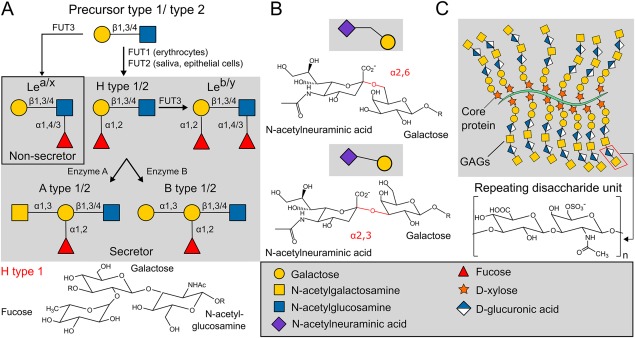
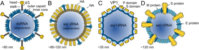
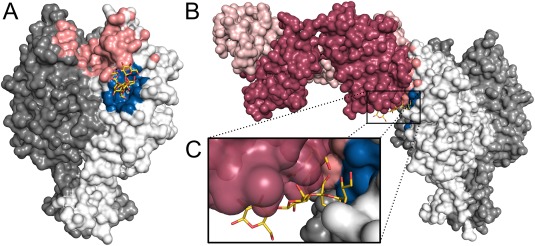
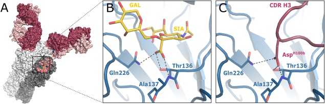
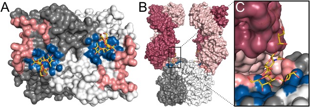
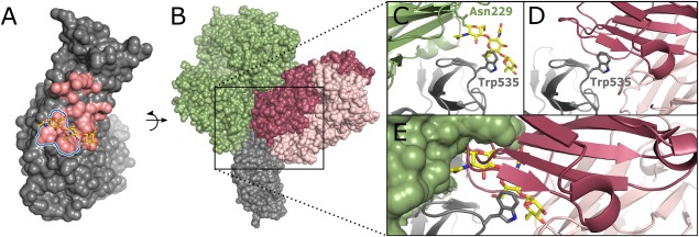
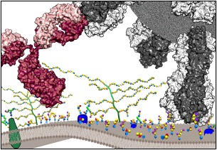
Numerous viruses rely on glycan receptor binding as the initial step in host cell infection. Engagement of specific glycan receptors such as sialylated carbohydrates, glycosaminoglycans, or histo-blood group antigens can determine host range, tissue tropism, and pathogenicity. Glycan receptor-binding sites are typically located in exposed regions on viral surfaces-sites that are also generally prone to binding of neutralizing antibodies that directly interfere with virus-glycan receptor interactions. In this review, we examine the locations and architecture of the glycan- and antibody-binding sites in four different viruses with stalk-like attachment proteins (reovirus, influenza virus, norovirus, and coronavirus) and investigate the mechanisms by which antibodies block glycan recognition. Those viruses exemplify that direct molecular mimicking of glycan receptors by antibodies is rare and further demonstrate that antibodies often partly overlap or bind sufficiently close to the receptor-binding region to hinder access to this site, achieving neutralization partially because of the epitope location and partly due to their sheer size.

Keywords: glycan receptors; neutralizing antibodies; structural characterization of binding epitopes and modes; viruses.


Figures

Figure 1
Figure 1
Glycan types that can function as viral receptors. (A) Biosynthesis of human ABH and Lewis HBGAs of Types 1 and 2. The types are defined by the glycosidic linkage of the precursor (Type 1 is β1,3 and Type 2 is β1,4 linked). Each step of the synthesis is catalyzed by a specific glycosyltransferase. FUT1 and FUT2 gene products control the same reaction. FUT1 is expressed in erythrocytes and FUT2 in secretory tissues giving rise to its glycosidic product in saliva and mucosal secretions. Sequential addition of monosaccharides to the precursor results in secretor‐HBGAs in the presence and to non‐secretor Lewis types in absence of FUT2 in secretions. FUT3 is primarily expressed in the epithelial cells of gastrointestinal tissue and adds a fucose to the precursor or H‐type antigens. Enzyme A or enzyme B adds GalNAc or Gal via α1,3 linkages to H‐type antigens, respectively, resulting in A and B type HBGAs. As an example H type 1 is shown in a structural representation. (B) Sialic acid variants. Sialic acids terminate N‐ and O‐glycans as well as glycolipids. The two common types of linkages, the α2,6‐ and α2,3‐ linkage, are shown with the most prominent sialic acid in humans, N‐acetylneuraminic acid, and Gal in a structural and schematic representation. The glycosidic linkage is highlighted in red. (C) In general GAGs are composed of repeating identical disaccharide units of N‐acetylated or N‐sulfated amino sugar linked to uronic acid or Gal. These units form long, unbranched GAG chains connected to a core protein. Depicted is chondroitin sulfate, a sulfated GAG consisting of repeating GalNAc and glucuronic acid units.
Figure 2
Figure 2
Morphology of viruses that contain spike‐like viral attachment proteins and are discussed in this review. (A) Mammalian reovirus contains a segmented double‐stranded (ds) RNA genome surrounded by two protein layers (inner core, outer capsid). The trimeric attachment protein σ1 is anchored into the capsid at the icosahedral vertices. Type 1 reoviruses engage sialylated carbohydrate receptors through the globular head domain (yellow) of σ1. (B) Influenza virus contains eight segments of single‐stranded (ss) RNA. The external layer contains the envelope glycoproteins HA and NA in an approximate ratio of 4:1. The HA spike is a homotrimer, whereas NA forms a tetramer. Although HA is responsible for binding sialylated glycans, NA is a receptor‐destroying enzyme that facilitates virus budding. (C) The norovirus particle is formed by 90 dimers of major capsid protein VP1 and encapsidates a (ss) RNA genome. The shell domain (S, in darker blue) of VP1 is involved in capsid formation, while the protruding domain (P, with subdomains P1 and P2 shown in light blue and yellow, respectively) projects from the shell surface at the icosahedral two‐fold axes. The P domain plays an important role in immune recognition and also binds to HBGAs. (D) CoVs contains a linear (ss) RNA genome. The viral membrane is comprised of membrane (M) and envelope small membrane (E) proteins (shown in light and dark blue, respectively). The trimeric spike proteins (S, highlighted in yellow) project from the surface and harbor RBSs. Due to the high sequence diversity among the S protein of CoV strains they bind to different receptors.
Figure 3
Figure 3
Binding of antibody 5C6 blocks glycan receptor engagement of the T1L reovirus protein σ1. Superposition of the T1L σ1 head domains of GM2 glycan (PDB ID: 4GU3) and Fab 5C6 (PDB ID: 5MHS) complex structures. (A) Surface representation of the trimeric σ1 head with monomers colored white, light and dark gray. The footprints of Fab 5C6 (salmon) and GM2 glycan (blue) binding have been calculated using a 4.5 Å distance cutoff. The carbohydrate molecules are shown as yellow sticks. (B) Side view of the σ1 head with one 5C6 Fab (light and heavy chain colored in light and dark violet, respectively). In this superposition, the Fab would clash with glycan moieties. For clarity, only one Fab and GM2 glycan are shown. (C) The close‐up view shows that there is not enough space for simultaneous binding of 5C6 and the glycan receptor.
Figure 4
Figure 4
Structures of influenza virus HA and binding interface of glycan receptor (PDB ID: 3UBJ) and antibody 5J8 (PDB ID: 5UGY). (A) Surface representation of influenza virus Cali07/2009 HA (one monomer of the trimeric molecule is shown in darker gray) with the binding epitope of the 5J8 antibody shown in salmon and the overlapping α2,6‐sialoglycan receptorbinding epitope in blue (cutoff of 4.5 Å). The sialoglycan is shown in stick representation with carbons in yellow. Two of the three bound Fabs are depicted in surface representation. (B) Detailed view of the binding interface of influenza virus Cali07/2009 HA with sialoglycan receptor. The sialoglycan receptor is shown in yellow as stick representation. Interactions of the sialic acid and the HA protein residues Gln226, Ala137, and Thr136 (also shown as sticks) are marked as black dashes. (C) Detailed view of the binding interface of influenza virus Cali07/2009 HA with the heavy chain CDR 3. The sialoglycan receptor mimicking residue AspH100b is depicted in stick representation. The residue engages the same residues as the sialoglycan receptor.
Figure 5
Figure 5
Binding of neutralizing antibody 5I2 to the Norovirus GI.1 P domain sterically hinders receptor HBGA binding. Superposition of the P domain complexed with HBGA (PDB ID: 2ZL6) and the P domain bound to Fab 5I2 (PDB ID: 5KW9). (A) Surface representation of the dimeric P domain with monomers colored white and gray. The epitope of Fab 5I2 (salmon) and the binding site of H‐type HBGA (blue) are highlighted (distance cutoff 4.5 Å). The two binding sites are close to each other, but different P domain residues are involved in the interactions. The glycan moieties are shown as yellow sticks. Due to crystal contacts that prevent ligand binding, one H‐type HBGA is modeled based on a superposition with the other subunit. (B) Side view of the P domain. Fabs 5I2 with light and heavy chain (colored in light and dark violet, respectively) binding their antigen‐ this engagement would partiallly mask and sterically block glycan binding. (C) The close up view shows a direct clash of glycan moieties and the heavy chain of 5I2.
Figure 6
Figure 6
Structures of MERS‐CoV RBD and binding surface of receptor DPP4 (PDB ID: 4KR0) and antibody MERS‐27 (PDB ID: 4ZS6). (A) Surface representation of MERS‐CoV RBD with the binding epitope of Fab MERS‐27 shown in salmon and the overlapping DPP4 carbohydrate‐binding epitope in blue (distance cutoff of 4.5 Å). The carbohydrate moiety is shown in stick representation, with carbons colored yellow. (B) Surface representation of MERS‐CoV RBD with DPP4 shown in green and Fab MERS‐27 with heavy and light chain (dark and light purple, respectively). Detailed view of the MERS‐CoV RBD with focus on residue Trp535. (C) DPP4 receptor binding on RBD (shown in green and gray cartoon representation, respectively) with the Asn229‐linked carbohydrate moiety in DPP4 (shown in yellow stick representation) interacting with residue Trp535. (D) Complex of RBD with MERS‐27 Fab. The heavy and light chain are colored in dark and light purple, respectively, and the central Trp535 residue is depicted in stick representation. (E) Representation of steric clashes upon simultaneous receptor and antibody binding. Since the binding site of the carbohydrate moiety completely overlaps with the Fab‐binding site, simultaneous binding is sterically not feasible.
Figure 7
Figure 7
Schematic representation of the competition between antibodies and glycan receptors for virus binding. Exemplified is a virus with trimeric spike proteins shown in gray and an antibody with heavy and light chain in dark and light purple, respectively. Cell‐surface carbohydrates (built with web‐based glycan modeler tools of: Woods Group [2005–2017] GLYCAM Web. Complex Carbohydrate Research Center, University of Georgia, Athens, GA. [http://glycam.org]) are shown in a schematic representation and are roughly scaled to the size of the viral spikes and antibody.

Similar articles

Cited by

References

    1. Varki NM, Varki A (2007) Diversity in cell surface sialic acid presentations: implications for biology and disease. Lab Invest 87:851–857. - PMC - PubMed
    1. Varki A, Schauer R (2009) Sialic acids In: Varki A, Cummings RD, Esko JD, Freeze HH, Stanley P, Bertozzi CR, Hart GW, Etzler ME, Eds. Essentials of Glycobiology, 2nd ed NY: Cold Spring Harbor. - PubMed
    1. Varki A, Gagneux P (2012) Multifarious roles of sialic acids in immunity. Ann N Y Acad Sci 1253:16–36. - PMC - PubMed
    1. Esko JD, Kimata K, Lindahl U (2009) Proteoglycans and sulfated glycosaminoglycans In: Varki A, Cummings RD, Esko JD, Freeze HH, Stanley P, Bertozzi CR, Hart GW, Etzler ME, Eds. Essentials of Glycobiology, 2nd ed NY: Cold Spring Harbor. - PubMed
    1. Marionneau S, Cailleau‐Thomas A, Rocher J, Le Moullac‐Vaidye B, Ruvoen N, Clement M, Le Pendu J (2001) ABH and Lewis histo‐blood group antigens, a model for the meaning of oligosaccharide diversity in the face of a changing world. Biochimie 83:565–573. - PubMed

LinkOut - more resources