

TREM2 expression in the human brain: a marker of monocyte recruitment?
- PMID: 28987033
- PMCID: PMC6221091
- DOI: 10.1111/bpa.12564
TREM2 expression in the human brain: a marker of monocyte recruitment?
Abstract
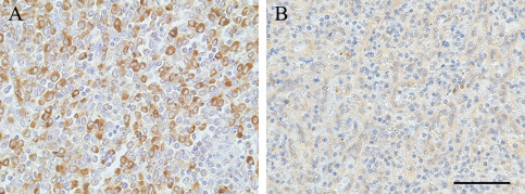
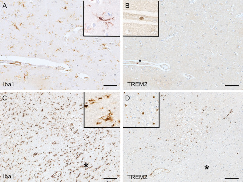
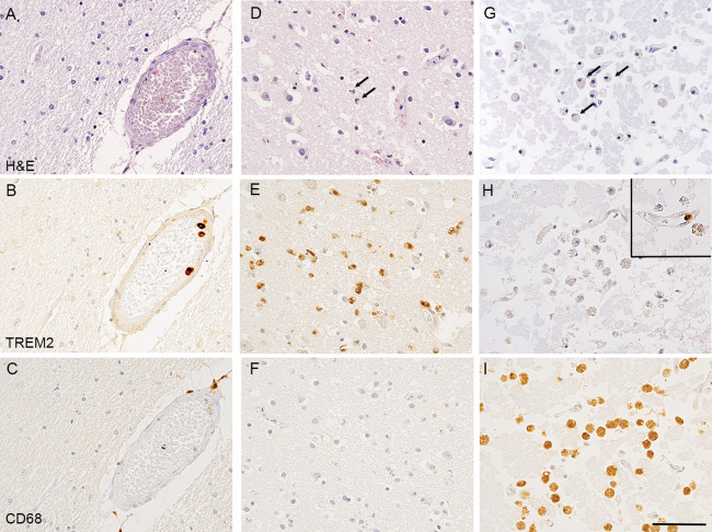
Mutation in the triggering receptor expressed on myeloid cells (TREM) 2 gene has been identified as a risk factor for several neurodegenerative diseases including Alzheimer's disease (AD). Experimental studies using animal models of AD have highlighted a number of functions associated with TREM2 and its expression by microglial cells. It has therefore been assumed that this is also the case in humans. However, there is very limited information concerning the cellular expression of TREM2 in the human brain. As part of investigations of microglia using post-mortem resources provided by the Medical Research Council Cognitive Function and Ageing Studies (MRC-CFAS), we immunostained the cerebral cortex of 299 participants for TREM2 using the Sigma antibody HPA010917 and compared with the macrophage/microglial markers Iba1 and CD68. As expected, Iba1 and CD68 labeled microglia and perivascular macrophages. However, in most cases (284/299), the TREM2 antibody labelled monocytes within vascular lumens, but not microglia or perivascular macrophages. In contrast, in 5 out of 6 cases with acute infarcts, TREM2 immunoreaction identified cells within the brain parenchyma interpreted as recruited monocytes. Six cases with old infarcts contained phagocytic foamy macrophages which were CD68-positive but TREM2 negative. Our observations, using the HPA010917 anti-TREM2 antibody, suggest that TREM2 is not expressed by microglia but instead seems to be a marker of recruited monocytes in the human brain. This finding has implications with regards to the role of TREM2 as a risk factor, emphasizing the importance of systemic immune responses in the development and progression of Alzheimer's disease.
Keywords: TREM2; dementia; human brain; microglia; monocyte; neuropathology.
© 2017 The Authors. Brain Pathology published by John Wiley & Sons Ltd on behalf of International Society of Neuropathology.
Conflict of interest statement
The authors do not have conflict of interest.
Figures
References
-
- Andersson PB, Perry VH, Gordon S (1991) The kinetics and morphological characteristics of the macrophage‐microglial response to kainic acid‐induced neuronal degeneration. Neuroscience 42:201–214. - PubMed
-
- Brayne C, McCracken C, Matthews FE, Medical Research Council Cognitive F , Ageing S (2006) Cohort profile: the Medical Research Council Cognitive Function and Ageing Study (CFAS). Int J Epidemiol 35:1140–1145. - PubMed
Publication types
MeSH terms
Substances
Grants and funding
- G0701018/MRC_/Medical Research Council/United Kingdom
- DH_/Department of Health/United Kingdom
- Walton Centre NHS Foundation Trust/International
- G0900652/MRC_/Medical Research Council/United Kingdom
- G0601022/MRC_/Medical Research Council/United Kingdom
- G0502157/MRC_/Medical Research Council/United Kingdom
- NIHR Cambridge Biomedical Research Centre/International
- G0400074/MRC_/Medical Research Council/United Kingdom
- UK NIHR Biomedical Research Centre/International
- G9901400/MRC_/Medical Research Council/United Kingdom
- MR/L022656/1/MRC_/Medical Research Council/United Kingdom
- University of Sheffield and the Sheffield Teaching Hospitals NHS Foundation Trust/International
- The Cambridgeshire and Peterborough NIHR CLAHRC/International
- G0900582/MRC_/Medical Research Council/United Kingdom
- G1100578/MRC_/Medical Research Council/United Kingdom
- Nottingham University Hospitals NHS Trust/International
- G1100540/MRC_/Medical Research Council/United Kingdom
- DH_/Department of Health/United Kingdom
- Oxford Biomedical Research Centre/International
- MR/N004272/1/MRC_/Medical Research Council/United Kingdom
LinkOut - more resources
Full Text Sources
Other Literature Sources
Research Materials
