Review
. 2017:145:263-283.
doi: 10.1016/B978-0-12-802395-2.00019-5.

Inflammatory demyelinating diseases of the central nervous system

Affiliations
Review

Inflammatory demyelinating diseases of the central nervous system

Romana Höftberger et al. Handb Clin Neurol. 2017.

Abstract

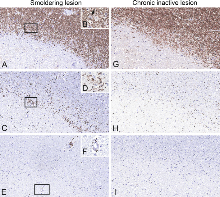
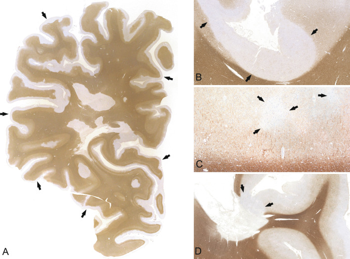
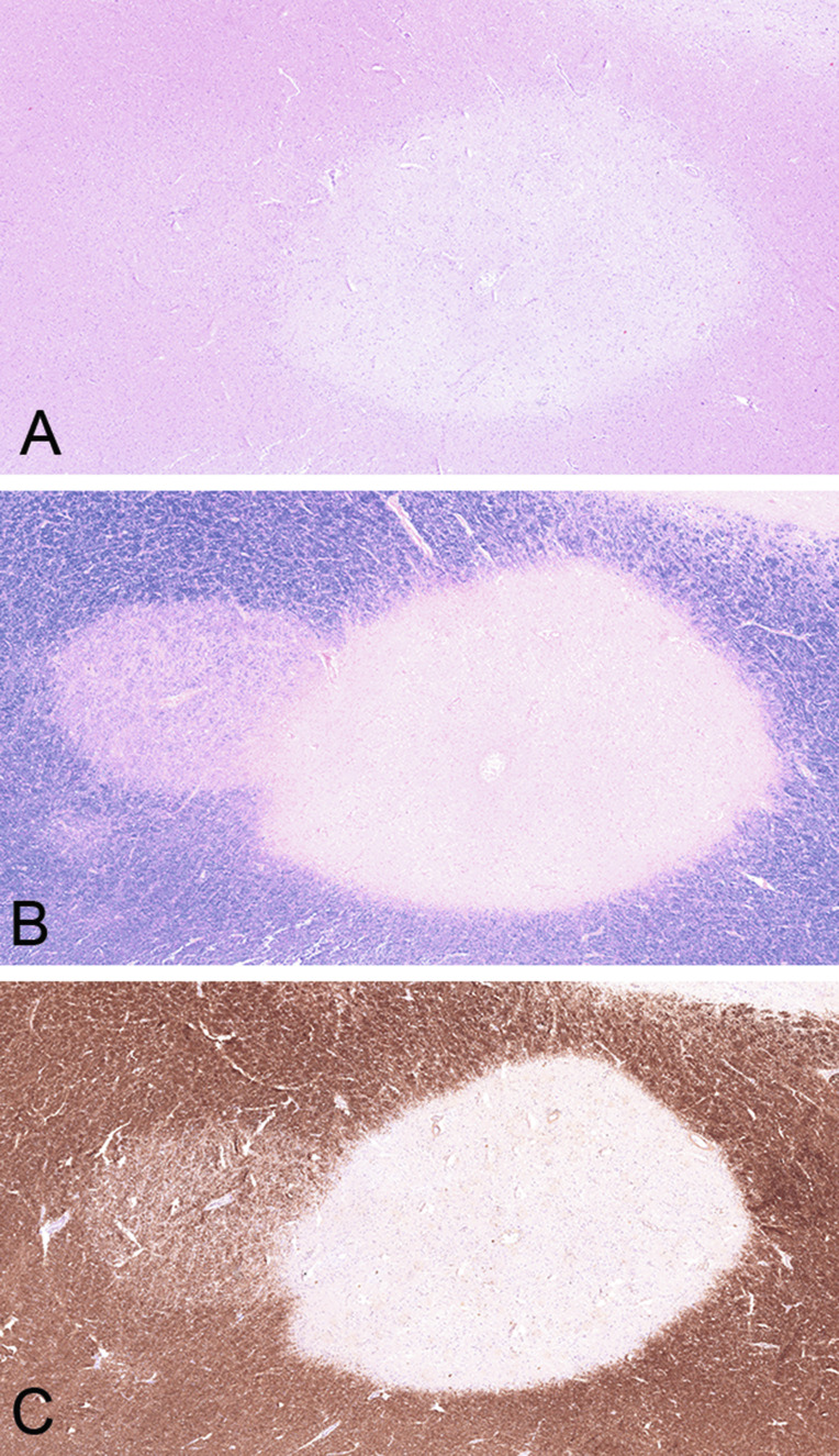
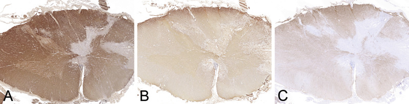
Inflammatory demyelinating diseases are a heterogeneous group of disorders, which occur against the background of an acute or chronic inflammatory process. The pathologic hallmark of multiple sclerosis (MS) is the presence of focal demyelinated lesions with partial axonal preservation and reactive astrogliosis. Demyelinated plaques are present in the white as well as gray matter, such as the cerebral or cerebellar cortex and brainstem nuclei. Activity of the disease process is reflected by the presence of lesions with ongoing myelin destruction. Axonal and neuronal destruction in the lesions is a major substrate for permanent neurologic deficit in MS patients. The MS pathology is qualitatively similar in different disease stages, such as relapsing remitting MS or secondary or primary progressive MS, but the prevalence of different lesion types differs quantitatively. Acute MS and Balo's type of concentric sclerosis appear to be variants of classic MS. In contrast, neuromyelitis optica (NMO) and spectrum disorders (NMOSD) are inflammatory diseases with primary injury of astrocytes, mediated by aquaporin-4 antibodies. Finally, we discuss the histopathology of other inflammatory demyelinating diseases such as acute disseminated encephalomyelitis and myelin oligodendrocyte glycoprotein antibody-associated demyelination. Knowledge of the heterogenous immunopathology in demyelinating diseases is important, to understand the clinical presentation and disease course and to find the optimal treatment for an individual patient.

Keywords: MOG antibodies; acute disseminated encephalomyelitis; aquaporin-4 antibodies; human autoimmune encephalomyelitis; multiple sclerosis; neuromyelitis optica.


Figures

Fig. 19.1
Fig. 19.1
Time course of myelin degradation in multiple sclerosis. Minor myelin proteins such as myelin oligodendrocyte glycoprotein (MOG) are degraded within 1–2 days of phagocytosis (A, arrows: MOG-positive myelin debris within macrophages), while major myelin proteins such as myelin basic protein (MBP) may persist for up to 6 days (B, arrows: MBP-positive myelin debris within macrophages). In later stages, macrophages contain periodic acid–Schiff (PAS)-positive residual glycoproteins (C, arrows: PAS-positive macrophages) and lipids (D, arrows: foamy and vacuolated macrophages). × 600.
Fig. 19.2
Fig. 19.2
Macroscopic appearance of multiple sclerosis. Chronic multiple sclerosis in a 61-year-old female with a 33-year history; section through the formalin-fixed brain (A) and the corresponding hemispheric section (B, luxol fast blue) shows multiple well-demarcated demyelinated plaques in the periventricular white matter (arrows).
Fig. 19.3
Fig. 19.3
Macroscopic appearance of multiple sclerosis in the brainstem. Chronic multiple sclerosis lesions (arrows) in the pons (A) and medulla oblongata (B) of a 47-year-old female with an 18-year history. Sections through the formalin-fixed brain (A, B) and corresponding histologic sections (C, D, luxol fast blue) show multiple well-demarcated demyelinated plaques (arrows).
Fig. 19.4
Fig. 19.4
Actively demyelinating lesion. Serial sections of the edge of an actively demyelinating multiple sclerosis plaque from a white-matter biopsy of a 22-year-old female. Periplaque white matter is seen on the right side of (A) and (B) (arrows). The plaque on the left side shows active demyelination with numerous macrophages containing myelin basic protein-positive myelin debris within their cytoplasm (A, rectangle enlarged upper right). Numerous CD68-positive macrophages (B) are mixed with CD3 + (C), CD4 + (D) and CD8 + parenchymal T cells (E) and perivenous CD79a + B cells/plasma cells (F, arrow). × 200.
Fig. 19.5
Fig. 19.5
Smoldering (slowly expanding) and inactive lesions. Serial sections of the edge of a smoldering demyelinating lesion (A, rectangle enlarged in B; myelin basic protein (MBP)) shows a rim of activated microglia and macrophages at the edge (C, rectangle enlarged in D; CD68). Few macrophages are digesting myelin components (B, arrow: macrophage with MPB-positive degradation products) and are accompanied by scattered CD8 + T cells (E, rectangle enlarged in F), suggesting an ongoing expansion into the surrounding normal-appearing white matter. In contrast, inactive lesions show thin myelin sheaths at the edge, representing remyelination (G), and only contain few microglia/macrophages (H) and T cells (I) at the edge. A, C, F, G–I, × 10; B, D, F, × 400.
Fig. 19.6
Fig. 19.6
Cortical demyelination. Cortical demyelination located subpially (A, B), intracortically (C), and overlapping with the white matter (so-called compound plaques) (D). A subpial lesion extends over several gyri and sulci and covers almost the entire cortex (A, proteolipid protein, arrows; B, myelin basic protein (MBP), arrows). A small, demyelinated lesion is located intracortically (C, MBP, arrows). A compound plaque is overlapping the cortex and the subcortical white matter (D, MBP, arrows). B, × 1.56; C, × 40; D, × 0.59.
Fig. 19.7
Fig. 19.7
Remyelinating shadow plaque. Overlap of a sharply demarcated chronic inactive plaque centered by a small vein, next to a remyelinated shadow plaque on the left, characterized by thinly myelinated fibers, demonstraded in hematoxylin and eosin (A), luxol fast blue (B), and myelin basic protein (C).
Fig. 19.8
Fig. 19.8
Spinal cord lesion in neuromyelitis optica. Destructive demyelinating lesion in the spinal cord (A, myelin basic protein) shows loss of astrocytes (B, aquaporin-4; C, aquaporin-1) and overlaps with the gray matter. × 1.1.
Fig. 19.9
Fig. 19.9
Active stage of neuromyelitis optica (NMO) lesion. An active NMO lesion shows inflammatory infiltrates with eosinophils (A, hematoxylin and eosin) next to macrophages and lymphocytes, deposition of immunoglobulin G (B, IgG), and C9 neoantigen (C, arrows). Some lesions show selective loss of aquaporin-4 (D, asterisk marks lesion) while aquaporin-1 is well preserved (E) and there is accumulation of bizarre astrocytes with beading and clumping of cell processes (clasmatodendrosis) (F, glial fibrillary acidic protein). A, B, F, × 600; C, × 200; D, E, × 100.
Fig. 19.10
Fig. 19.10
Vacuolated lesions in neuromyelitis optica (NMO). A vacuolated lesion in NMO is characterized by expansion of the extracellular space and shows some apoptotic nuclei (A, B, hematoxylin and eosin, arrow: apoptotic cell). The myelin is relatively well preserved with only few demyelinating macrophages (C, D, luxol fast blue (LFB); arrow: macrophage with LFB-positive degradation products; E, F, myelin basic protein; arrowhead: apoptotic cell), in contrast, astrocytes are lost (G, H, glial fibrillary acid protein). B, D, F, H: × 250.
Fig. 19.11
Fig. 19.11
Acute disseminated encephalomyelitis (ADEM). Lesions in ADEM are characterized by small perivenous demyelinating areas (A, luxol fast blue (LFB)), with numerous macrophages containing LFB-positive degradation products (B, LFB; C, CD68). A, C, × 100; B, × 600.
Fig. 19.12
Fig. 19.12
Myelin oligodendrocyte glycoprotein (MOG) antibody-associated demyelinating encephalomyelitis. A demyelinating lesion associated with MOG antibodies is well demarcated (A, luxol fast blue) and shows relatively well-preserved (B, CNPase; C, MOG) oligodendrocytes and deposition of C9 neoantigen (D, arrows). × 200.

References

    1. Aboul-Enein F., Rauschka H., Kornek B. Preferential loss of myelin-associated glycoprotein reflects hypoxia-like white matter damage in stroke and inflammatory brain diseases. J Neuropathol Exp Neurol. 2003;62:25–33. - PubMed
    1. Aloisi F., Pujol-Borrell R. Lymphoid neogenesis in chronic inflammatory diseases. Nature reviews. Immunology. 2006;6:205–217. - PubMed
    1. Anlar B., Basaran C., Kose G. Acute disseminated encephalomyelitis in children: outcome and prognosis. Neuropediatrics. 2003;34:194–199. - PubMed
    1. Babbe H., Roers A., Waisman A. Clonal expansions of CD8(+) T cells dominate the T cell infiltrate in active multiple sclerosis lesions as shown by micromanipulation and single cell polymerase chain reaction. J Exp Med. 2000;192:393–404. - PMC - PubMed
    1. Balo J. Encephalitis periaxialis concentrica. Arch Neurol Psychiatry. 1928;19:242–264.

MeSH terms