Review
. 2017;38(2):255-276.

Pathophysiology of the Effects of Alcohol Abuse on the Endocrine System

Affiliations
Free PMC article
Review

Pathophysiology of the Effects of Alcohol Abuse on the Endocrine System

Nadia Rachdaoui et al. Alcohol Res. 2017.
Free PMC article

Abstract

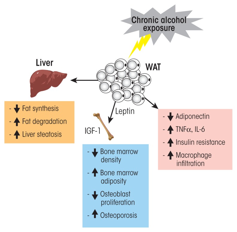
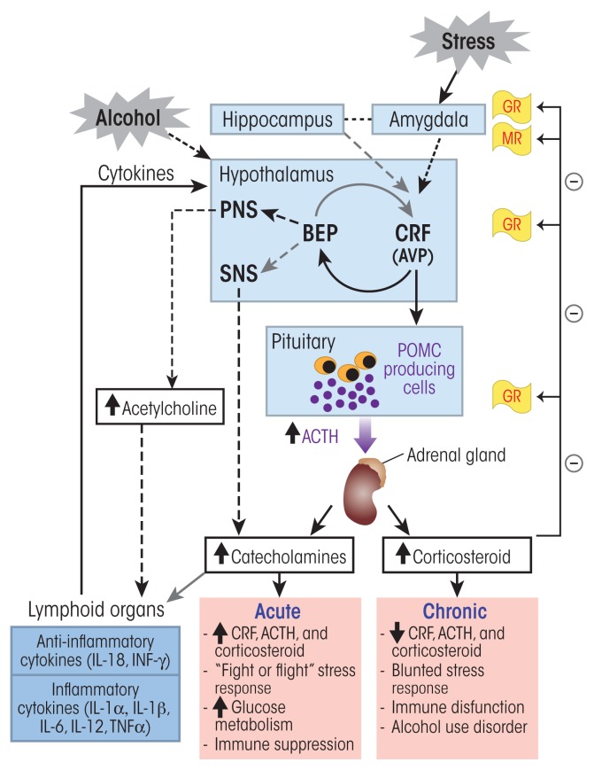
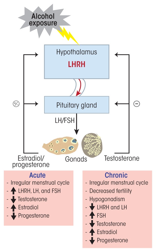
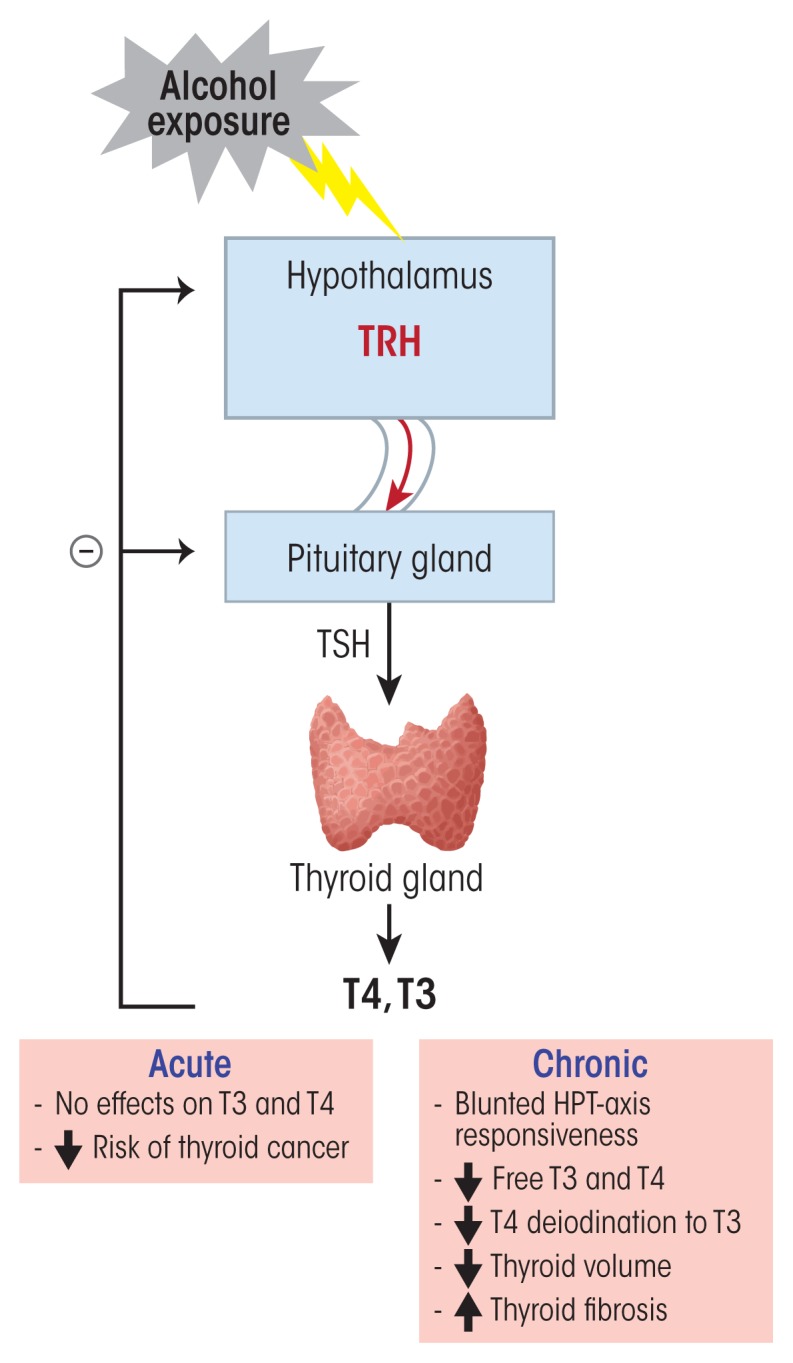
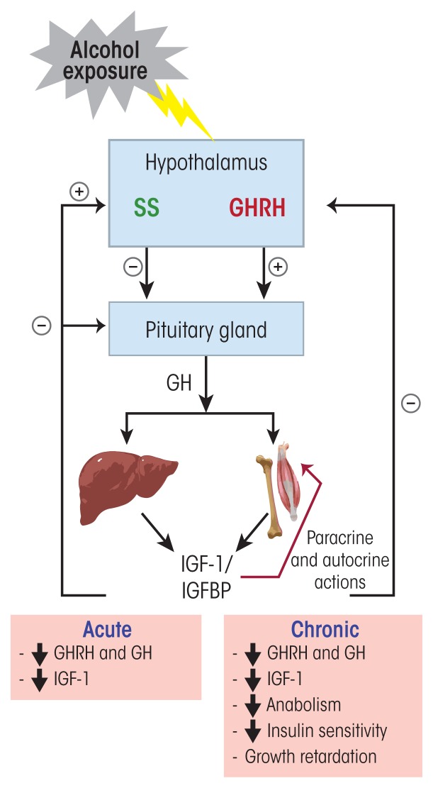
Alcohol can permeate virtually every organ and tissue in the body, resulting in tissue injury and organ dysfunction. Considerable evidence indicates that alcohol abuse results in clinical abnormalities of one of the body's most important systems, the endocrine system. This system ensures proper communication between various organs, also interfacing with the immune and nervous systems, and is essential for maintaining a constant internal environment. The endocrine system includes the hypothalamic-pituitary-adrenal axis, the hypothalamic-pituitary-gonadal axis, the hypothalamic-pituitary-thyroid axis, the hypothalamic-pituitary-growth hormone/insulin-like growth factor-1 axis, and the hypothalamic-posterior pituitary axis, as well as other sources of hormones, such as the endocrine pancreas and endocrine adipose tissue. Alcohol abuse disrupts all of these systems and causes hormonal disturbances that may result in various disorders, such as stress intolerance, reproductive dysfunction, thyroid problems, immune abnormalities, and psychological and behavioral disorders. Studies in both humans and animal models have helped shed light on alcohol's effects on various components of the endocrine system and their consequences.


Conflict of interest statement

Financial Disclosure

The authors declare that they have no competing financial interests.

Figures

None
Alcohol and the endocrine white adipose tissue (WAT). WAT is a dynamically active endocrine organ that produces and secretes adipokines, including hormones, growth factors, and cytokines. These factors, through autocrine, paracrine, and endocrine actions, can influence the function of many tissues and coordinate numerous important biological processes such as food intake, glucose homeostasis, lipid metabolism, and pro- and anti-inflammatory functions. Acute and moderate alcohol exposure induces an increase in circulating adiponectin levels, which is associated with decreased insulin resistance. Chronic alcohol exposure induces a decrease in adiponectin, an increase in macrophage infiltration and proinflammatory cytokine secretion (e.g., tumor necrosis factor alpha (TNFα) and interleukin-6 [IL-6]) and insulin resistance. Chronic alcohol exposure also increases the risk of fatty liver (i.e., steatosis).
Figure 1
Figure 1
Alcohol’s effects on the hypothalamic–pituitary–adrenal (HPA) axis and the stress response. Alcohol can stimulate neurons in the paraventricular nucleus (PVN) of the hypothalamus to release corticotropin-releasing factor (CRF) and arginine vasopressin (AVP). Stress sensed in the amygdala also elicits a similar activation of this stress response pathway. In the anterior pituitary, CRF stimulates the production of proopiomelanocortin (POMC), which serves as the prohormone for adrenocorticotropic hormone (ACTH). AVP potentiates the effects of CRF on ACTH release from the anterior pituitary. ACTH stimulates cells of the cortical portion of adrenal glands to produce and release glucocorticoid hormones (i.e., cortisol). High levels of glucocorticoids inhibit CRF and ACTH release through a negative feedback by binding to glucocortiocoid receptors (GRs) and mineralocorticoid receptors (MRs) in various brain regions. Neurons in the arcuate nucleus of the hypothalamus release β-endorphin (BEP), which also regulates CRF release. BEP also acts on the autonomous nervous system and inhibits the sympathetic nervous system (SNS) stress response. CRF, ACTH, and glucocorticoids also act on different organs of the immune system and stimulate cytokine production and release into the general circulation. These cytokines then reach the brain where they trigger a neuroimmune response that sensitizes the stress-response pathway. Acute exposure to alcohol stimulates the HPA-axis stress response and induces suppression of cytokine production. In contrast, chronic exposure to alcohol induces a blunted HPA-axis stress response characterized by an absence of negative feedback control of this pathway and an increase in proinflammatory cytokines, such as interleukin-6 (IL-6) and tumor necrosis factor alpha (TNFα), leading to stress intolerance, immune dysfunction and alcohol use disorder.
Figure 2
Figure 2
Alcohol’s effects on the hypothalamic–pituitary–gonadal (HPG) axis. Neurons in the hypothalamus release luteinizing hormone–releasing hormone (LHRH) to the hypophyseal-portal blood system. LHRH then stimulates the secretion of gonadotropins (i.e., LH and FSH). During the ovary’s follicular phase, FSH stimulates the development of a dominant follicle, which produces and secretes estradiol. Estradiol then stimulates an LH and FSH surge during midcycle of the menstrual cycle. LH stimulates ovulation and the development of the corpus luteum, which then produces and secretes progesterone. In the testis, LH stimulates testosterone production and release, while FSH controls spermatogenesis. HPG axis function is controlled through feedback loop mechanisms. Testosterone inhibits LHRH, LH, and FSH secretion through negative feedback, whereas estradiol and progesterone both can have negative- and positive-feedback actions, depending on the stage of the ovarian cycle, and can inhibit or stimulate the release of LHRH, LH, and FSH. Acute alcohol exposure results in increased LHRH, LH, FSH, and estradiol and decreased testosterone and progesterone. Chronic alcohol exposure, in contrast, induces a decrease in LHRH, LH, testosterone, and progesterone and an increase in estradiol and FSH. These alcohol-induced hormonal dysregulations cause a multitude of reproductive disorders, such as menstrual cycle irregularity, decreased fertility, and hypogonadism.
Figure 3
Figure 3
Alcohol’s effects on the hypothalamic–pituitary–thyroid (HPT) axis. Thyrotropin-releasing hormone (TRH) released from neurons in the hypothalamus stimulates thyrotropic cells in the anterior pituitary to produce and secrete thyroid-stimulating hormone (TSH). TSH then stimulates the synthesis and secretion of thyroxin (T4) and its active form, triiodothyronine (T3), from the follicular cells of the thyroid gland. Circulating T3 comes from conversion of T4 by enzymes called deiodinases in the liver. T3 and T4 can control their own release by negative feedback at the hypothalamus and the pituitary and inhibit TRH and TSH release. Acute alcohol exposure has no effect on HPT-axis function. However, chronic alcohol exposure leads to a blunted TSH response to TRH, as well as to decreased free T3 and T4, decreased deiodination of T4 to T3, decreased thyroid volume, and increased thyroid fibrosis.
Figure 4
Figure 4
Alcohol’s effects on the growth hormone–insulin-like growth factor-1 (GH/IGF-1) axis. Growth hormone (GH)-releasing hormone (GHRH) secreted from neurons in the hypothalamus acts on somatotropic cells in the anterior pituitary and stimulates the production and release of GH into the circulation. GH can act on target tissues and directly affect their function or it can stimulate IGF-1 production and secretion from these target tissues, especially from the liver. IGF-1 then is either released into the general circulation, where it circulates bound to IGF binding proteins (IGFBP), or it can mediate GH anabolic effects on target tissues through paracrine and autocrine actions. Through negative feedback at the hypothalamus and pituitary, IGF-1 can reduce GHRH and GH secretion. Somatostatin (SS), secreted in the paraventricular nucleus of the hypothalamus, also acts on the pituitary and inhibits GH secretion. IGF-1 stimulates SS secretion. Acute and chronic alcohol exposure leads to decreased GHRH, GH, and IGF-1 secretion.

References

    1. Ben-Jonathan N, Hnasko R. Dopamine as a prolactin (PRL) inhibitor. Endocrine Reviews. 2001;22(6):724–763. - PubMed
    1. Chaturvedi K, Sarkar DK. Alteration in G proteins and prolactin levels in pituitary after ethanol and estrogen treatment. Alcoholism: Clinical and Experimental Research. 2008;32(5):806–813. - PMC - PubMed
    1. De A, Boyadjieva N, Oomizu S, Sarkar DK. Ethanol induces hyperprolactinemia by increasing prolactin release and lactotrope growth in female rats. Alcoholism: Clinical and Experimental Research. 2002;26(9):1420–1429. - PubMed
    1. De A, Boyadjieva N, Pastorcic M, Sarkar D. Potentiation of the mitogenic effect of estrogen on the pituitary-gland by alcohol-consumption. International Journal of Oncology. 1995;7(3):643–648. - PubMed
    1. Dees WL, Kozlowski GP. Differential effects of ethanol on luteinizing hormone, follicle stimulating hormone and prolactin secretion in the female rat. Alcohol. 1984;1(6):429–433. - PubMed

Publication types

MeSH terms