Review
. 2018 Jan:114:40-51.
doi: 10.1016/j.freeradbiomed.2017.10.001. Epub 2017 Oct 6.

Dysfunction of autophagy and endosomal-lysosomal pathways: Roles in pathogenesis of Down syndrome and Alzheimer's Disease

Affiliations
Review

Dysfunction of autophagy and endosomal-lysosomal pathways: Roles in pathogenesis of Down syndrome and Alzheimer's Disease

Daniel J Colacurcio et al. Free Radic Biol Med. 2018 Jan.

Abstract

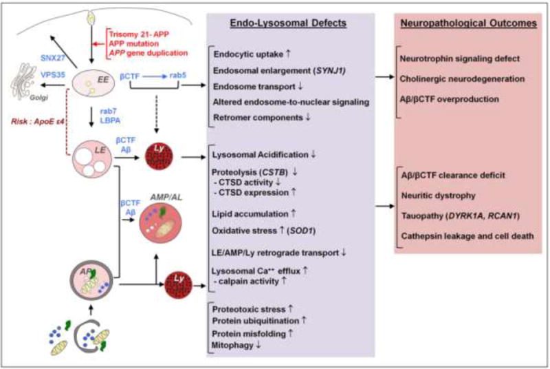
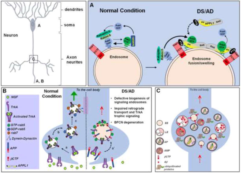
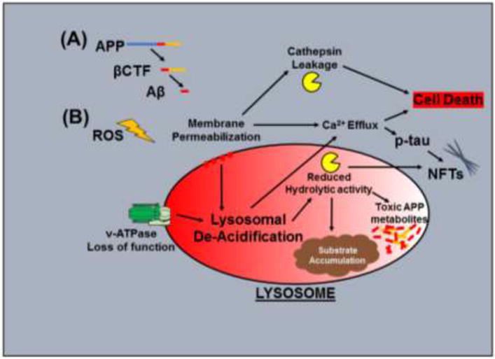
Individuals with Down syndrome (DS) have an increased risk of early-onset Alzheimer's Disease (AD), largely owing to a triplication of the APP gene, located on chromosome 21. In DS and AD, defects in endocytosis and lysosomal function appear at the earliest stages of disease development and progress to widespread failure of intraneuronal waste clearance, neuritic dystrophy and neuronal cell death. The same genetic factors that cause or increase AD risk are also direct causes of endosomal-lysosomal dysfunction, underscoring the essential partnership between this dysfunction and APP metabolites in AD pathogenesis. The appearance of APP-dependent endosome anomalies in DS beginning in infancy and evolving into the full range of AD-related endosomal-lysosomal deficits provides a unique opportunity to characterize the earliest pathobiology of AD preceding the classical neuropathological hallmarks. Facilitating this characterization is the authentic recapitulation of this endosomal pathobiology in peripheral cells from people with DS and in trisomy mouse models. Here, we review current research on endocytic-lysosomal dysfunction in DS and AD, the emerging importance of APP/βCTF in initiating this dysfunction, and the potential roles of additional trisomy 21 genes in accelerating endosomal-lysosomal impairment in DS. Collectively, these studies underscore the growing value of investigating DS to probe the biological origins of AD as well as to understand and ameliorate the developmental disability of DS.

Keywords: Alzheimer's Disease; Autophagy; Down syndrome; Endosomes; Lysosomes.


Figures

Figure 1
Figure 1
Overview of the shared pathways and pathological mechanisms driven by the APP gene and its cleavage products (βCTF/Aβ) in DS and AD: known and predicted endocytic and lysosomal factors contributing to neurodegeneration. Genetic contributors to neurodegeneration in DS other than APP are shown in parentheses. ApoE ε4, a known genetic risk factor for AD, contributes to endolysosomal dysfunction in AD and potentially in DS. EE, early endosome; LE, late endosome; AP, autophagosome; Ly, Lysosome; AMP/AL, amphisome/autolysosome.
Figure 2
Figure 2
Endosomal pathology and altered endosome trafficking in DS and AD contribute to aberrant neurotrophin transport and signaling and neuritic dystrophy. A) Under normal conditions, the conversion of rab5 from an active to an inactive state occurs rapidly and it is highly regulated by the coordinated action of rab5 effectors rabex-5/rabaptin-5 and rabGAP5, respectively. In DS and AD, APP-βCTF mediates pathological activation of rab5 by recruiting APPL1 to endosomes. APPL1 binds to the “YENPT” motif of βCTF through its phosphotyrosine binding (PTB) domain. Upon dimerization via its BAR domains to facilitate vesicle curvature, APPL1 binds to GTP-rab5 via its PH domain, which stabilizes rab5 in its activated state on endosomes. Over-activation of rab5 causes acceleration of endocytosis and endosome fusion leading to characteristic swelling of endosomes. B) Under normal conditions, biogenesis of long-lived signaling endosomes involves endocytosis of activated TrkA receptors (NGF-bound TrkA) into rab5-positive endosomes (a), followed by deactivation of rab5 and acquisition of rab7 (b), which binds to dynein/dynactin motor complexes regulating long-range retrograde axonal transport (c, green arrow). In DS and AD, prolonged rab5 hyperactivation by APP/APP-βCTF and APPL1 causes enlargement of NGF/TrkA endosomes (a’) and slows retrograde endosome transport (red dotted arrow), thus diminishing TrkA signaling which ultimately leads to the loss of trophic support for basal forebrain cholinergic neurons (BFCNs). C) DS and AD are characterized by the pathological accumulation of ubiquitinated misfolded/aggregated proteins and of enlarged early and late endosomal structures (EE and LE, respectively) containing βCTF/Aβ fragments. Autophagosomes (AP), amphisomes (AMP), and endolysosomal compartments containing incompletely degraded material accumulate within neuritic swellings (“dystrophic neurites”) as a result of the βCTF/Aβ-mediated impairment of retrograde transport and lysosomal function.
Figure 3
Figure 3
Common mechanisms of lysosomal dysfunction in neurons during AD and DS: (A) APP and its cleavage products, βCTF and Aβ, contribute to lysosomal de-acidification and lysosomal membrane permeabilization, leading to a decline in lysosomal function. (B) Reactive oxygen species (ROS), which are increased in DS and in AD, also contribute to impaired lysosomal function. Congruent elements of lysosomal dysfunction; including lysosomal de-acidification, reduced proteolysis, lysosomal membrane permeabilization leading to loss of membrane integrity, and the release of Ca2+ and cathepsins represent common pathological outcomes in AD and DS, leading to accumulation of intralysosomal waste, toxic APP and tau metabolite accumulation, neurofibrillary tangle (NFT) formation, and cell death.

References

    1. Arai Y, Ijuin T, Takenawa T, Becker LE, Takashima S. Excessive expression of synaptojanin in brains with Down syndrome. Brain Dev. 2002;24:67–72. - PubMed
    1. Ascano M, Richmond A, Borden P, Kuruvilla R. Axonal targeting of Trk receptors via transcytosis regulates sensitivity to neurotrophin responses. J Neurosci. 2009;29:11674–11685. - PMC - PubMed
    1. Avrahami L, Farfara D, Shaham-Kol M, Vassar R, Frenkel D, Eldar-Finkelman H. Inhibition of glycogen synthase kinase-3 ameliorates beta-amyloid pathology and restores lysosomal acidification and mammalian target of rapamycin activity in the Alzheimer disease mouse model: in vivo and in vitro studies. J Biol Chem. 2013;288:1295–1306. - PMC - PubMed
    1. Babst M. MVB vesicle formation: ESCRT-dependent, ESCRT-independent and everything in between. Curr Opin Cell Biol. 2011;23:452–457. - PMC - PubMed
    1. Bhaskar K, Miller M, Chludzinski A, Herrup K, Zagorski M, Lamb BT. The PI3K-Akt-mTOR pathway regulates Abeta oligomer induced neuronal cell cycle events. Mol Neurodegener. 2009;4:14. - PMC - PubMed

Publication types