. 2017 Nov 7;26(5):764-777.e5.
doi: 10.1016/j.cmet.2017.09.002. Epub 2017 Oct 5.

Lipolysis in Brown Adipocytes Is Not Essential for Cold-Induced Thermogenesis in Mice

Affiliations

Lipolysis in Brown Adipocytes Is Not Essential for Cold-Induced Thermogenesis in Mice

Hyunsu Shin et al. Cell Metab. .

Abstract

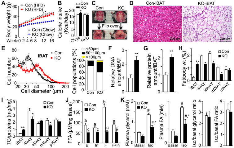
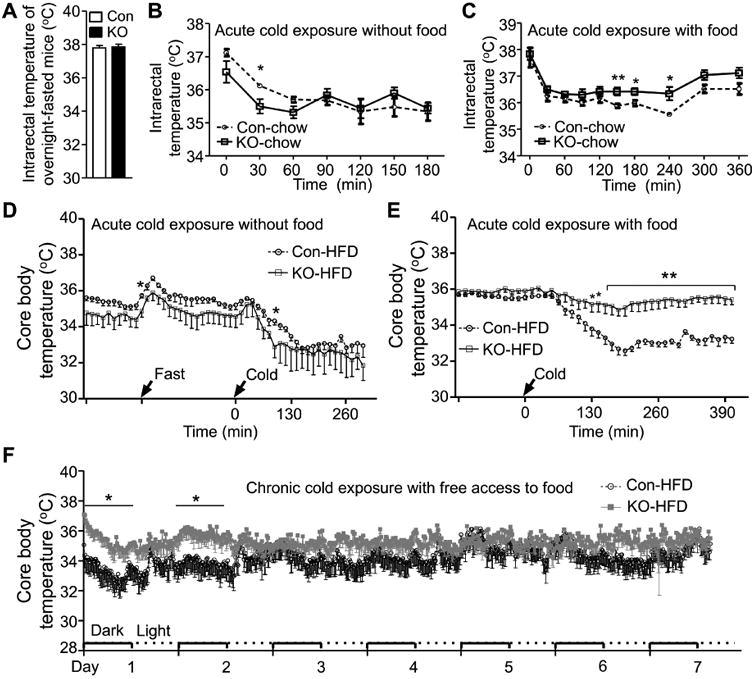
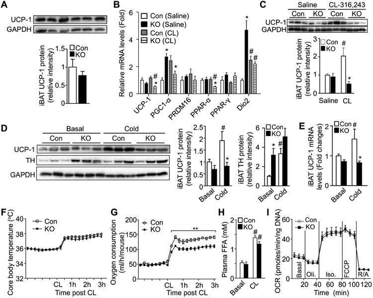
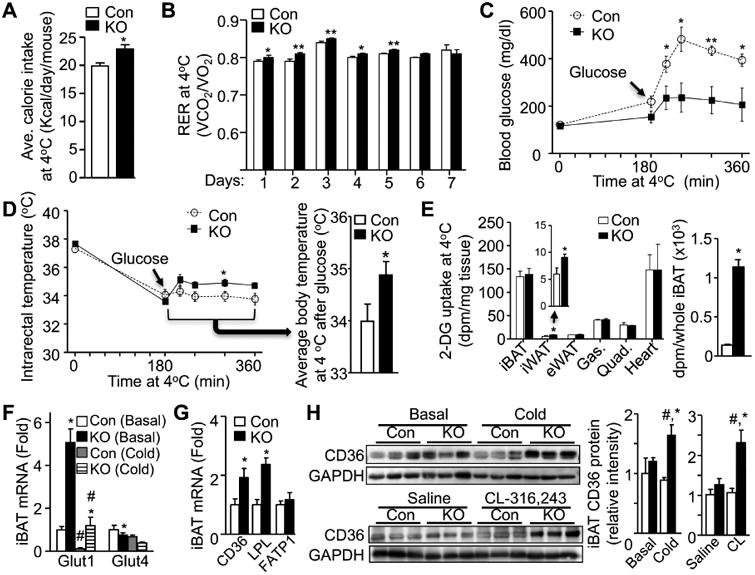
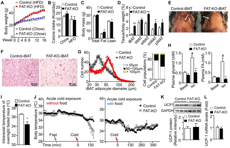
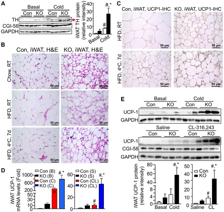
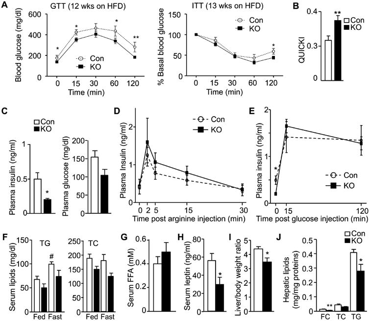
Lipid droplet (LD) lipolysis in brown adipose tissue (BAT) is generally considered to be required for cold-induced nonshivering thermogenesis. Here, we show that mice lacking BAT Comparative Gene Identification-58 (CGI-58), a lipolytic activator essential for the stimulated LD lipolysis, have normal thermogenic capacity and are not cold sensitive. Relative to littermate controls, these animals had higher body temperatures when they were provided food during cold exposure. The increase in body temperature in the fed, cold-exposed knockout mice was associated with increased energy expenditure and with increased sympathetic innervation and browning of white adipose tissue (WAT). Mice lacking CGI-58 in both BAT and WAT were cold sensitive, but only in the fasted state. Thus, LD lipolysis in BAT is not essential for cold-induced nonshivering thermogenesis in vivo. Rather, CGI-58-dependent LD lipolysis in BAT regulates WAT thermogenesis, and our data uncover an essential role of WAT lipolysis in fueling thermogenesis during fasting.

Keywords: CGI-58; beige adipocyte; body temperature; energy expenditure; intracellular lipolysis; metabolic health; sympathetic activation; thermogenesis; white adipose tissue browning.


Figures

Figure 1
Figure 1. Selective Inactivation of CGI-58 in BAT Induces Steatotic Hypertrophy and Hyperplasia and Blocks the Stimulated Lipolysis in iBAT
The data in this figure were obtained from the mice housed at room temperature (22°C). (A) Weight gain of 6-week-old (Week 0) BAT-KO (KO) and control (Con) mice subjected to chow (n = 10/group) or HFD (n = 11/group). (B) Food intake of mice on the fifth week of chow (n = 6/group) or HFD (n = 11/group). (C) Gross appearance of iBATs in 24-week-old mice on chow diet. (D) H&E staining of iBATs from 14-week-old chow-fed mice. (E) Average brown adipocyte sizes in iBATs of mice on HFD for 18 weeks. For each mouse, 5,000 cells were measured. n = 5/group. (F, G) Relative DNA and protein amounts per iBAT in 14-week-old mice on chow. n = 5/group. (H) Adipose depot/body weight ratios (%) of mice on HFD for 5 weeks. n =7/group. (I) The TG content in individual adipose depots of mice on HFD for 18 weeks. n = 5-7/group. (J) Lipolysis assays of iBAT explants from 28-day-old mice. n = 5/group. B, basal; In, ATGL inhibitor Atglistatin; F, forskolin. Statistically significant differences are indicated by different small letters (p < 0.05, One-way ANOVA). (K) In vivo lipolysis capacity of mice on HFD for 4 weeks. n = 5/group.
Figure 2
Figure 2. BAT-KO Mice Are Not Cold Sensitive
(A) Intrarectal temperatures of 20-week-old HFD-fed BAT-KO (KO) and control (Con) mice at 22°C. n = 4-5/group. (B, C) Intrarectal temperatures of 10-11-week-old chow-fed mice exposed 4°C in the absence or presence of diet. n = 5/group. (D) Core body temperatures of 28-week-old HFD-fed mice exposed to 4°C in the absence of food. n = 5-6/group. (E) Core body temperatures of 24-week-old HFD-fed mice exposed to 4°C in the presence of food. n = 5-6/group. (F) Core body temperatures of 18-week-old HFD-fed mice during 7 days of cold exposure. n = 6/group.
Figure 3
Figure 3. CGI-58 Deletion in BAT Does Not Reduce Whole-body Thermogenic Capacity
(A) Western blots and densitometry of iBAT UCP1 protein in 14-week-old chow-fed mice. (B) Relative iBAT expression levels of thermogenesis-related genes from the mice injected with CL316,243 for 4 days (CL) or vehicle (Saline). n = 5/group. (C) Western blots and densitometry of iBAT UCP1 in the same mice described under 3B. (D) Western blots and densitometry analyses of iBAT UCP1 and TH proteins in 18-week-old HFD-fed mice at 22°C (Basal) or 4°C for 7 days (Cold). (E) UCP1 mRNA levels in the iBAT of the mice described under 3D. n = 4-5/group. (F and G) Core body temperatures (F) and oxygen consumption rates (G) in 23-week-old mice injected with a bolus of CL316,243 at thermoneutrality. n = 6/group. (H) Plasma levels of FFAs in thermoneutrally housed 11-week-old mice at 15 min after a bolus of CL316,243 injection. n = 6-7/group. (I) Oxygen consumption rates (OCRs) measured by Seahorse assays in isolated primary brown adipocytes from 28-day-old mice. Oli., oligomycin; Iso., isoproterenol; R/A, rotenone/antimycin.
Figure 4
Figure 4. BAT-KO Mice Increase Combustion of Circulating Fuels During Cold Exposure
(A and B) Average daily calorie intake (A) and respiratory exchange ratio (RER) (B) of 18-week-old HFD-fed BAT-KO (KO) and control (Con) mice during 7 days of cold exposure. n = 5-6/group. (C and D) Changes in blood glucose levels (C) and intrarectal temperatures (D) in 12-week-old HFD-fed mice after a bolus of glucose injection (i.p., at 1.5 g/kg body weight). The mice were fasted for 7h (4h at room temperature and 3h at cold) prior to glucose injection. n = 4-5/group. (E) Tissue glucose uptake in 20-week-old HFD-fed mice during acute cold exposure. The total glucose uptake of iBAT was calculated based on the total iBAT weight. n = 4-5/group. (F) Levels of mRNAs for glucose transporter 1 (Glut1) and glucose transporter 4 (Glut4) in iBAT in mice housed at 22°C (Basal) or 4°C for 7 days (Cold). n = 5/group. (G) Levels of mRNAs for CD36, lipoprotein lipase (LPL), and fatty acid transporter protein 1 (FATP1) in iBAT in 14-week-old HFD-fed mice housed at 22°C. n = 5/group. (H) Immunoblots and densitometry of CD36 protein in iBAT in mice exposed to cold for 7 days or injected daily with CL316,243 for 4 days.
Figure 5
Figure 5. WAT Lipolysis Is Essential for Thermogenesis During Fasting
(A) Weight gain of FAT-KO and control mice on chow (n = 9-12) or HFD (n = 10-13. Mice were 6 weeks old at Week 0. (B) Calorie intake on the fifth week of chow (n = 5-6/group) or HFD (n = 5-6/group) treatment. (C) Body composition of 12-week-old HFD-fed mice. n = 6/group. (D) Adipose depot/body weight ratios in 24-week-old HFD-fed mice. n = 7/group. (E) Gross appearance of iBATs from 24-week-old HFD-fed mice. (F) H&E staining of iBATs from 14-week-old HFD-fed mice. (G) Adipocyte size of iBATs from 24-week-old HFD-fed mice. For each sample, 5,000 cells were measured. n = 5/group. (H) In vivo lipolysis capacity of 10-week-old HFD-fed mice. (I) Intrarectal temperatures of 26-week-old HFD-fed mice during cold exposure in the absence of food. n = 5-6/group. (J) Core body temperatures of 23-week-old HFD-fed mice during cold exposure in the absence or presence of food. n = 5-6/group. (K) Western blots and densitometry of UCP1 protein in iBAT in 22-week-old HFD-fed mice. (L) Levels of UCP1 mRNA in iBAT in 22-week-old HFD-fed mice. n = 4/group.
Figure 6
Figure 6. CGI-58 Deletion in BAT Stimulates WAT Browning
(A) Western blots and densitometry of tyrosine hydroxylase (TH) in iWAT in 18-week-old HFD-fed mice exposed to cold for 7 days. (B) H&E staining of iWATs from 14-week-old chow-fed BAT-KO (KO) and control (Con) mice (top), 18-week-old HFD-fed mice (middle), and 18-week-old HFD-fed mice exposed to cold for 7 days (bottom). (C) Immunostaining of UCP1 protein in iWAT in 18-week-old HFD-fed mice or 18-week-old HFD-fed mice exposed to cold for 7 days. (D and E) Levels of UCP1 mRNA (D) and protein (E) in iWAT in 18-week-old HFD-fed mice at 22°C (Basal, or B) or 4°C for 7 days (Cold, or C) (n = 4-5/group), or 14-week-old HFD-fed mice injected with CL316,243 for 4 days (CL) or saline (S) (n = 5/group).
Figure 7
Figure 7. CGI-58 Deletion in BAT Improves Metabolic Profile in Mice
(A) Glucose tolerance test (GTT) and insulin tolerance test (ITT) in BAT-KO (KO) and control (Con) mice on HFD. n = 9-11/group. (B) QUICKI insulin sensitivity indexes of overnight-fasted 17-week-old HFD-fed mice. N = 6/group. (C) Plasma concentrations of insulin and glucose in 17-week-old HFD-fed mice fasted overnight. n = 6/group. (D) Arginine-stimulated insulin secretion in 8-week-old mice. n = 6/group. (E) Glucose-stimulated insulin secretion in 17-week-old HFD-fed mice. n = 6/group. (F) Serum concentrations of triglycerides (TG) and total cholesterol (TC) in the fed or overnight-fasted 15-17-week-old HFD-fed mice. n = 6/group. (G) Serum concentrations of FFAs in 17-week-old HFD-fed mice fasted overnight. n = 6/group. (H) Serum concentrations of leptin in 15-week-old HFD-fed mice in the fed state. n = 6/group. (I) Liver/body weight ratios in 24-week-old HFD-fed mice. n = 7/group. (J) Hepatic contents of free cholesterol (FC), total cholesterol (TC), and TG in 24-week-old HFD-fed mice. n = 5/group.

Comment in

  • What Ignites UCP1?
    Cannon B, Nedergaard J. Cannon B, et al. Cell Metab. 2017 Nov 7;26(5):697-698. doi: 10.1016/j.cmet.2017.10.012. Cell Metab. 2017. PMID: 29117542

References

    1. Ahmadian M, Abbott MJ, Tang T, Hudak CS, Kim Y, Bruss M, Hellerstein MK, Lee HY, Samuel VT, Shulman GI, et al. Desnutrin/ATGL is regulated by AMPK and is required for a brown adipose phenotype. Cell Metab. 2011;13:739–748. - PMC - PubMed
    1. Altshuler-Keylin S, Shinoda K, Hasegawa Y, Ikeda K, Hong H, Kang Q, Yang Y, Perera RM, Debnath J, Kajimura S. Beige Adipocyte Maintenance Is Regulated by Autophagy-Induced Mitochondrial Clearance. Cell Metab. 2016;24:402–419. - PMC - PubMed
    1. Bartelt A, Bruns OT, Reimer R, Hohenberg H, Ittrich H, Peldschus K, Kaul MG, Tromsdorf UI, Weller H, Waurisch C, et al. Brown adipose tissue activity controls triglyceride clearance. Nat Med. 2011;17:200–205. - PubMed
    1. Blondin DP, Frisch F, Phoenix S, Guerin B, Turcotte EE, Haman F, Richard D, Carpentier AC. Inhibition of Intracellular Triglyceride Lipolysis Suppresses Cold-Induced Brown Adipose Tissue Metabolism and Increases Shivering in Humans. Cell Metab. 2017;25:438–447. - PubMed
    1. Cannon B, Nedergaard J. Brown adipose tissue: function and physiological significance. Physiol Rev. 2004;84:277–359. - PubMed

Substances