

Structure of the Guanidine III Riboswitch
- PMID: 28988949
- PMCID: PMC5696562
- DOI: 10.1016/j.chembiol.2017.08.021
Structure of the Guanidine III Riboswitch
Abstract
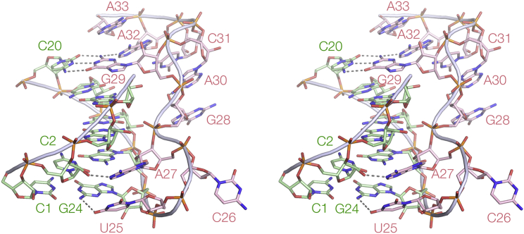
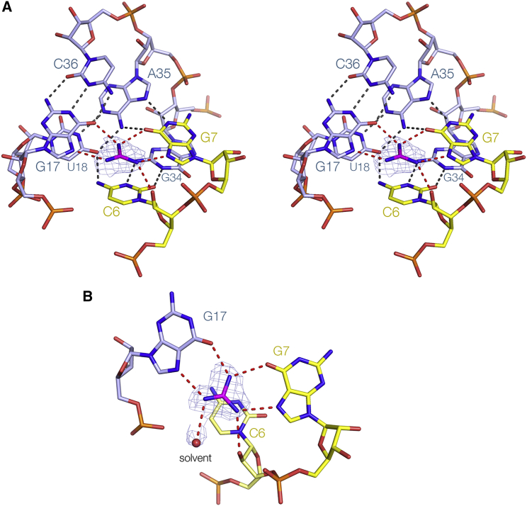
Riboswitches are structural elements found in mRNA molecules that couple small-molecule binding to regulation of gene expression, usually by controlling transcription or translation. We have determined high-resolution crystal structures of the ykkC guanidine III riboswitch from Thermobifida fusca. The riboswitch forms a classic H-type pseudoknot that includes a triple helix that is continuous with a central core of conserved nucleotides. These form a left-handed helical ramp of inter-nucleotide interactions, generating the guanidinium cation binding site. The ligand is hydrogen bonded to the Hoogsteen edges of two guanine bases. The binding pocket has a side opening that can accommodate a small side chain, shown by structures with bound methylguanidine, aminoguanidine, ethylguanidine, and agmatine. Comparison of the new structure with those of the guanidine I and II riboswitches reveals that evolution generated three different structural solutions for guanidine binding and subsequent gene regulation, although with some common elements.
Keywords: RNA structure; X-ray crystallography; gene regulation; riboregulation.
Copyright © 2017 The Authors. Published by Elsevier Ltd.. All rights reserved.
Figures


References
-
- Beaucage S.L., Caruthers M.H. Deoxynucleoside phosphoramidites – a new class of key intermediates for deoxypolynucleotide synthesis. Tetrahedron Lett. 1981;22:1859–1862.
MeSH terms
Substances
Grants and funding
LinkOut - more resources
Full Text Sources
Other Literature Sources
