. 2017 Oct:24:64-75.
doi: 10.1016/j.ebiom.2017.09.010. Epub 2017 Sep 13.

IRX3 Promotes the Browning of White Adipocytes and Its Rare Variants are Associated with Human Obesity Risk

Affiliations

IRX3 Promotes the Browning of White Adipocytes and Its Rare Variants are Associated with Human Obesity Risk

Yaoyu Zou et al. EBioMedicine. 2017 Oct.

Abstract

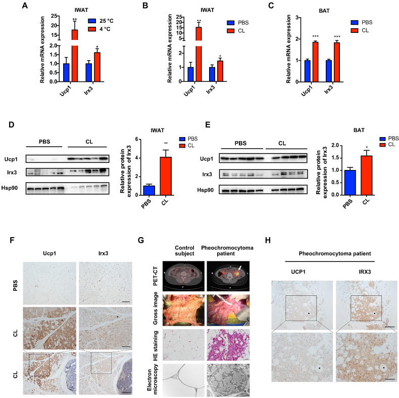
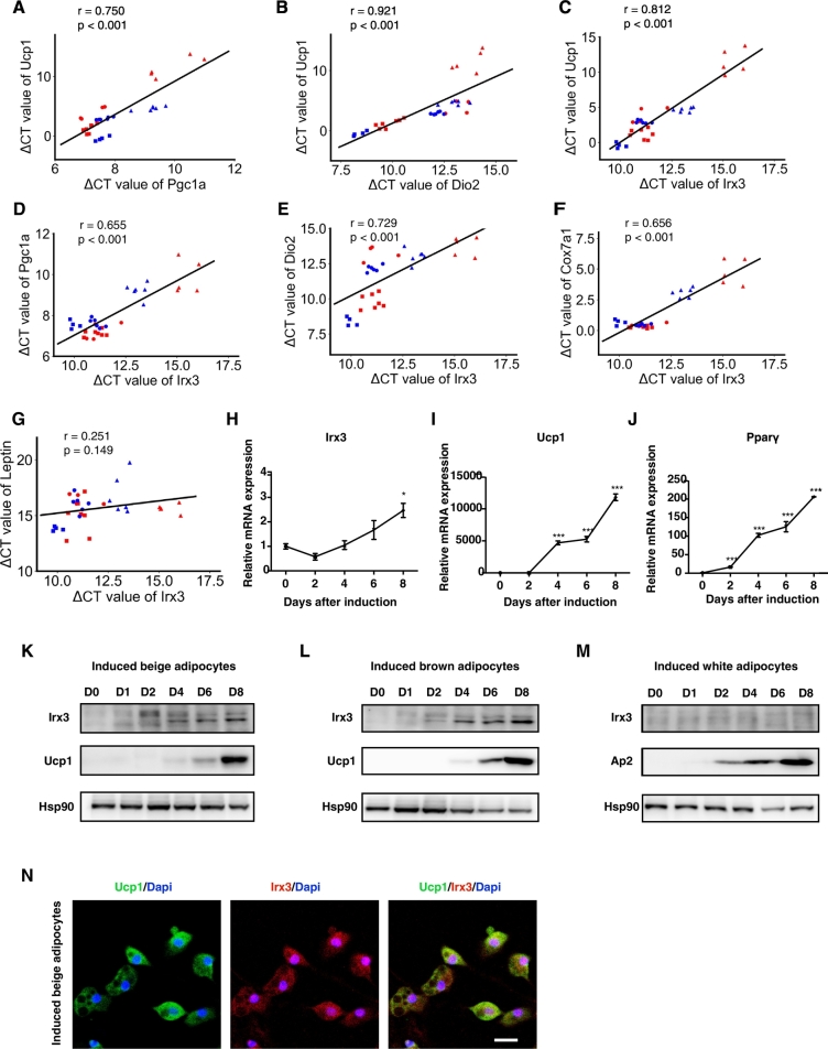
Background: IRX3 was recently reported as the effector of the FTO variants. We aimed to test IRX3's roles in the browning program and to evaluate the association between the genetic variants in IRX3 and human obesity.

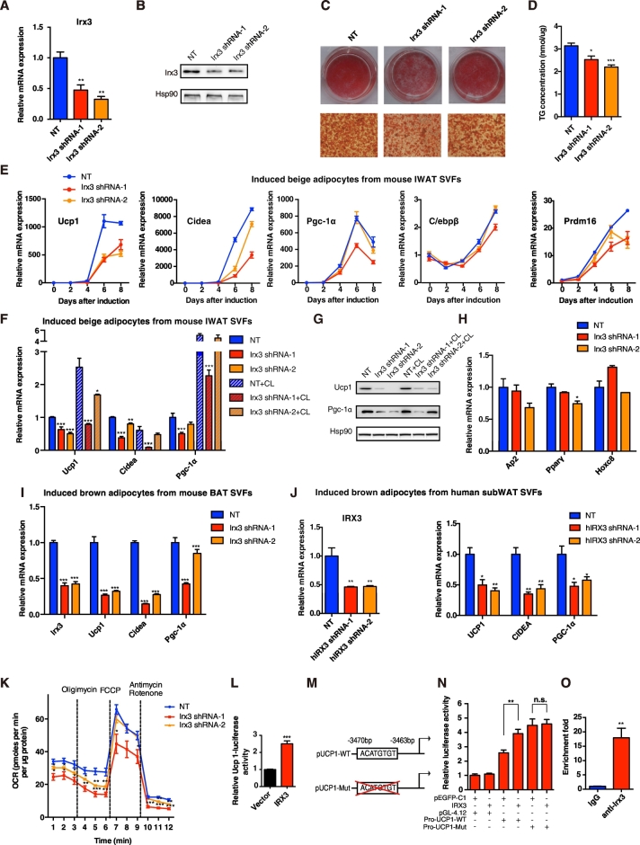
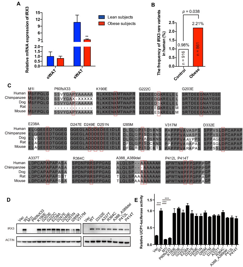
Methods: IRX3 expression was examined in beige adipocytes in human and mouse models, and further validated in induced beige adipocytes. The browning capacity of primary preadipocytes was assessed with IRX3 knockdown. Luciferase reporter analysis and ChIP assay were applied to investigate IRX3's effects on UCP1 transcriptional activity. Moreover, genetic analysis of IRX3 was performed in 861 young obese subjects and 916 controls.

Results: IRX3 expression was induced in the browning process and was positively correlated with the browning markers. IRX3 knockdown remarkably inhibited UCP1 expression in induced mouse and human beige adipocytes, and also repressed the uncoupled oxygen consumption rate. Further, IRX3 directly bound to UCP1 promoter and increased its transcriptional activity. Moreover, 17 rare heterozygous missense/frameshift IRX3 variants were identified, with a significant enrichment in obese subjects (P=0.038, OR=2.27; 95% CI, 1.02-5.05).

Conclusions: IRX3 deficiency repressed the browning program of white adipocytes partially by regulating UCP1 transcriptional activity. Rare variants of IRX3 were associated with human obesity.

Keywords: Beige adipocytes; Browning program; Genetic variants; IRX3; Obesity.


Figures

Fig. 1
Fig. 1
IRX3/Irx3 expression is induced in browning adipose tissues. (A) Relative mRNA levels of Ucp1 and Irx3 in IWAT from mice under cold stress (4 °C) for one week (n = 6) and at 25 °C (n = 8). (B–C) Relative mRNA levels of Ucp1 and Irx3 in IWAT (B) and BAT (C) from mice subjected to PBS or CL-316,243 for 10 days. For A–C, gene expression was normalized to 36b4. (D–E) Protein levels of Ucp1 and Irx3 (left) and the quantification value of Irx3 relative to Hsp90 (right) in IWAT (D) and BAT (E) in the mice subjected to PBS or CL-316,243 treatment. (F) Representative images of immunohistochemical staining in IWAT from mice subjected to PBS or CL-316,243 treatment (scale bar, 50 μm for the upper and middle panels, and 100 μm for the bottom panel). (G) PET-CT, gross image, HE staining (scale bar, 20 μm), and electron microscopy (scale bar, 5 μm) of the browning WAT from pheochromocytoma patients. (H) Representative images of immunohistochemical staining in oWAT from pheochromocytoma patients (scale bar, 100 μm for the upper panel, and 50 μm for the bottom panel). Data were presented as mean ± s.e.m. *P < 0.05, **P < 0.01, ***P < 0.001. The results are representative of at least three independent experiments. IWAT, inguinal white adipose tissue. BAT, brown adipose tissue.
Fig. 2
Fig. 2
The expression of Irx3 was correlated with the beige/brown adipocyte associated genes in vitro. (A–G) In fully differentiated beige/brown adipocytes from preadipocytes isolated from IWAT, EWAT, and BAT of C57BL/6J and 129/Sv mice, Ucp1 mRNA expression was correlated with Pgc-1α (A) and Dio2 (B). Irx3 mRNA expression showed positive correlation with mRNA levels of (C) Ucp1, (D) Pgc-1α, (E) Dio2, and (F) Cox7a1. Irx3 mRNA expression showed no correlation with Leptin (G). Cells from C57BL/6J were shown in red, and from 129/Sv were in blue. Cells from IWAT, EWAT, and BAT were shown as dot, triangle, and square, respectively. The correlation was analyzed using ΔCT. (H-J) (H) Irx3 (I) Ucp1 and (J) Pparγ mRNA expression during the time course of beige adipocyte differentiation cultures (n = 3). Quantitative amounts of gene expression were normalized to the housekeeping gene 36b4. (K–L) Irx3 and Ucp1 protein levels at different time points during beige adipocyte differentiation from IWAT SVFs (K), during brown adipocyte differentiation from BAT SVFs (L). (M) Protein expression of Irx3 and Ap2 at different time points during white adipocyte differentiation from IWAT SVFs. (N) Intracellular location of Ucp1 and Irx3 protein in the fully differentiated beige adipocytes. Ucp1 protein, Irx3 protein and nucleus were indicated in green, red, and blue, respectively. Scar bar, 20 μm. Data were presented as mean ± s.e.m. *P < 0.05, **P < 0.01, ***P < 0.001. The results are representative of at least three independent experiments.
Fig. 3
Fig. 3
Knockdown of Irx3 repressed the differentiation of SVFs toward beige adipocytes in vitro. (A–B) (A) Relative mRNA and (B) protein levels of Irx3 in SVFs from IWAT were efficiently knocked down after a 3-day infection of mouse Irx3 lentiviral shRNA (n = 3 for each group). (C–D) (C) Oil Red O staining and (D) TG concentration of the mature beige adipocytes under eight-day differentiation (n = 6 for each group). (E) Relative mRNA expression of Ucp1, Cidea, Pgc-1α, C/ebpβ, and Prdm16 at different time points during beige adipocyte differentiation from IWAT SVFs (n = 3–4 for different groups). (F–G) (F) Relative mRNA (n = 4) and (G) protein expression of brown adipocyte marker genes of the induced beige adipocytes from mouse IWAT SVTs for eight days with the infection of mouse Irx3 lentiviral shRNA, with or without CL-316,243 activation. (H) Relative mRNA expression of adipocyte differentiation-related genes in beige adipocytes (n = 4). (I) Relative mRNA expression of the indicated genes in the induced brown adipocytes from mouse BAT SVFs for eight days, with the infection of mouse Irx3 lentiviral shRNA (n = 4). (J) Relative mRNA levels of IRX3 in SVFs from human sWAT after a 3-day infection of human IRX3 lentiviral shRNA (left), and the mRNA levels of brown adipocyte marker genes in the SVFs under 2-day differentiation (n = 4). (K) OCR measurement of the beige adipocytes under 5-day differentiation, with the infection of mouse Irx3 lentiviral shRNA (n = 5). (L–N) (L) Ucp1 promoter-luciferase reporter activity was measured in HEK293T cells transfected with pEGFP-C1 vector or IRX3 for 48 h. (M) Schematic diagram of the mutant mouse Ucp1 promoter deleting ACATGTGT among the − 3470 to − 3463 bp region proximal to the TSS of the Ucp1 gene. (N) Transcriptional activity of wild-type or mutant Ucp1 promoter was measured with IRX3 overexpression. (O) qPCR of Ucp1 promoter binding region which was recruited by Irx3 antibody in 8-day induced beige adipocytes, as analyzed by ChIP. Fold enrichment of Ucp1 promoter was given (n = 3). For the measurement of luciferase activity, cells were seeded on 24-well plate and transfected with 800 ng pEGFP-C1 vector or IRX3, 200 ng Ucp1 promoter construct, and 1 ng pRL-SV40, followed by the harvest for luciferase activity assessment using a dual-luciferase reporter assay system (Promega). Luciferase activity was corrected for Renilla luciferase activity (n = 3–4 for each group). For qPCR data, mRNA expression was normalized to 36b4 for mouse genes and βACTIN for human genes. SVF cells were isolated from IWAT and BAT of C57BL/6J mice or human sWAT wherever mentioned, and IRX3 shRNA were introduced to cells 24 h after seeding. Data were presented as mean ± s.e.m. *P < 0.05, **P < 0.01, ***P < 0.001. The results are representative of at least three independent experiments. NT, no targeting.
Fig. 4
Fig. 4
The association of IRX3 and human obesity. (A) Relative mRNA levels of IRX3 in oWAT or sWAT from normal weight (n = 9) and obese subjects (n = 21) measured by qPCR. Gene expression was normalized to βACTIN. (B) Comparison of the frequency of the IRX3 rare variants in normal weight subjects and obese patients. (C) Sequence conservation analysis of the IRX3 orthologs related to variants identified in obese subjects and controls. Mutant sites are shown with a red box. Dark gray represents residues that are completely conserved, mild gray partially conserved, and light gray shows similar residues. (D) Protein expression levels of WT and mutant IRX3 in HEK293T cells. GFP antibody was used to indicate IRX3, and actin antibody served as a loading control. (E) Ucp1 promoter-luciferase reporter activity was measured in HEK293T cells transfected with pEGFP-C1 vector, WT or mutant IRX3 for 48 h (n = 3). Normalized luciferase activities are shown as fold change and compared to WT IRX3 (n = 4). oWAT, omental white adipose tissue. sWAT, subcutaneous white adipose tissue. Data were presented as mean ± s.e.m. *P < 0.05, **P < 0.01, ***P < 0.001. The results are representative of at least three independent experiments.

Comment in

Similar articles

Cited by

References

    1. Allison D.B., Kaprio J., Korkeila M., Koskenvuo M., Neale M.C., Hayakawa K. The heritability of body mass index among an international sample of monozygotic twins reared apart. Int. J. Obes. Relat. Metab. Disord. 1996;20:501–506. - PubMed
    1. Bellefroid E.J., Kobbe A., Gruss P., Pieler T., Gurdon J.B., Papalopulu N. Xiro3 encodes a Xenopus homolog of the Drosophila Iroquois genes and functions in neural specification. EMBO J. 1998;17:191–203. - PMC - PubMed
    1. Bilioni A., Craig G., Hill C., McNeill H. Iroquois transcription factors recognize a unique motif to mediate transcriptional repression in vivo. Proc. Natl. Acad. Sci. U. S. A. 2005;102:14671–14676. - PMC - PubMed
    1. Bonnefond A., Clément N., Fawcett K., Yengo L., Vaillant E., Guillaume J.L. Rare MTNR1B variants impairing melatonin receptor 1B function contribute to type 2 diabetes. Nat. Genet. 2012;44:297–301. - PMC - PubMed
    1. Claussnitzer M., Dankel S.N., Kim K.H., Quon G., Meuleman W., Haugen C. FTO obesity variant circuitry and adipocyte Browning in humans. N. Engl. J. Med. 2015;373:895–907. - PMC - PubMed

MeSH terms