

New diagnostic tools for breast cancer
- PMID: 28989543
- PMCID: PMC5605595
- DOI: 10.1007/s12254-017-0341-5
New diagnostic tools for breast cancer
Abstract
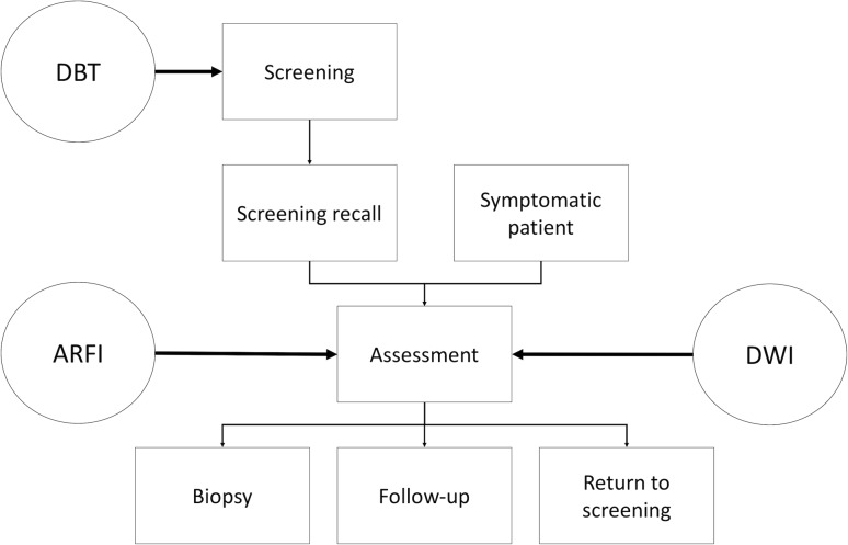
Imaging plays a major role in the diagnosis, treatment, and follow-up of breast cancer. Findings that require further assessment will be detected both at screening and curative mammography. Most findings that are further worked up tend to yield benign diagnoses. Consequently, there is an ongoing search for new tools to reduce recalls and unnecessary biopsies while maintaining or improving cancer detection rates. The clinically most promising methods in this respect are described and discussed in this review.
Keywords: Breast cancer; Contrast-enhanced mammography; Elastography; MRI; Tomosynthesis.
Conflict of interest statement
Conflict of interest
P.A.T. Baltzer, P. Kapetas, M.A. Marino, and P. Clauser declare that they have no competing interests.
Ethical standards
This chapter does not contain any studies with human participants or animals performed by any of the authors.
Figures
References
-
- Sardanelli F, Aase HS, Álvarez M, Azavedo E, Baarslag HJ, Balleyguier C, et al. Position paper on screening for breast cancer by the European Society of Breast Imaging (EUSOBI) and 30 national breast radiology bodies from Austria, Belgium, Bosnia and Herzegovina, Bulgaria, Croatia, Czech Republic, Denmark, Estonia, Finland, France, Germany, Greece, Hungary, Iceland, Ireland, Italy, Israel, Lithuania, Moldova, The Netherlands, Norway, Poland, Portugal, Romania, Serbia, Slovakia, Spain, Sweden, Switzerland and Turkey. Eur Radiol. 2016 - PMC - PubMed
Publication types
LinkOut - more resources
Full Text Sources
Other Literature Sources