. 2017 Aug 1;8(8):5769-5780.
doi: 10.1039/c7sc01765k. Epub 2017 Jun 6.

Stimuli-responsive nucleic acid-functionalized metal-organic framework nanoparticles using pH- and metal-ion-dependent DNAzymes as locks

Affiliations

Stimuli-responsive nucleic acid-functionalized metal-organic framework nanoparticles using pH- and metal-ion-dependent DNAzymes as locks

Wei-Hai Chen et al. Chem Sci. .

Abstract

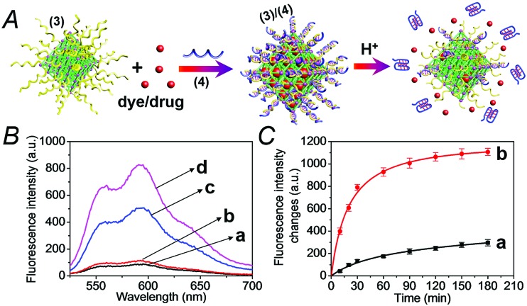
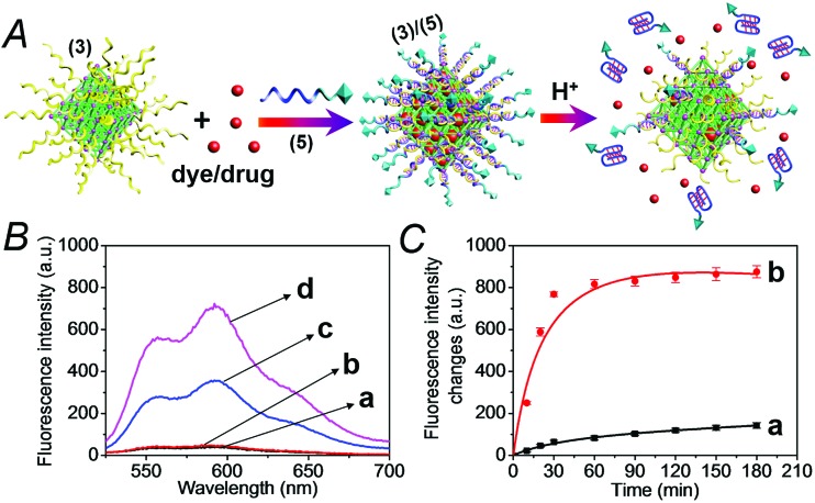
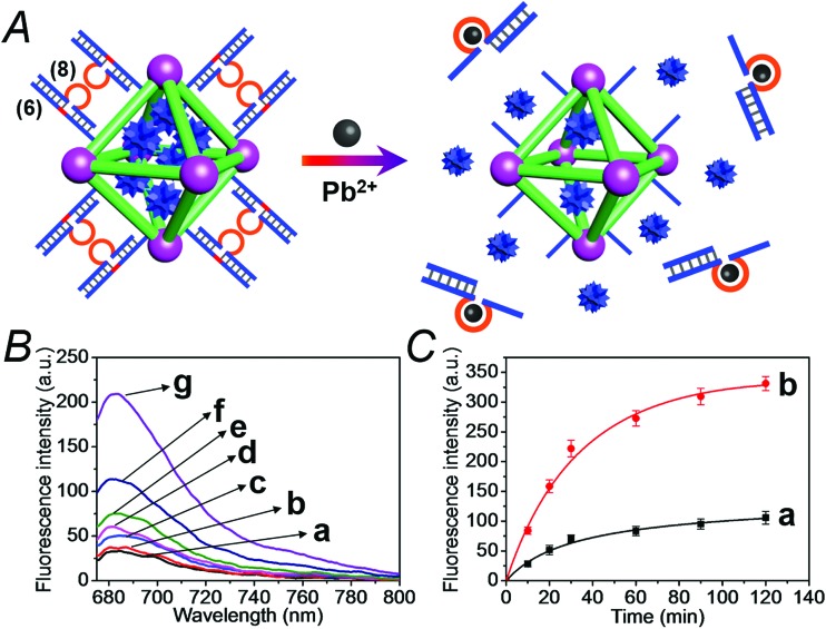
A versatile approach to modify metal-organic framework nanoparticles (NMOFs) with nucleic acid tethers, using the "click chemistry" method is introduced. The nucleic acid-functionalized NMOFs are used to prepare stimuli-responsive carriers of loads (fluorescence probes or anti-cancer drugs). Two different stimuli-responsive nucleic acid-based NMOFs are presented. One system involves the preparation of pH-responsive NMOFs. The NMOFs are loaded with fluorophores or doxorubicin anti-cancer drug and locked in the NMOFs by pH-responsive DNA duplex capping units. At pH = 5.0 the capping units are unlocked, leading to the release of the loads. The AS1411 aptamer is conjugated to the locking units as the targeting unit for the nucleolin biomarker present in cancer cells. The pH-responsive doxorubicin-loaded NMOFs and, in particular, the AS1411 aptamer-modified pH-responsive NMOFs reveal selective, targeted, cytotoxicity toward MDA-MB-231 breast cancer cells. A second system involves the synthesis of NMOFs that are loaded with fluorophores or doxorubicin and capped with metal-ion-dependent DNAzyme/substrate complexes as locking units (metal ion = Mg2+ or Pb2+ ions). In the presence of the respective metal ions, the nucleic acid locking units are cleaved off, resulting in the release of the loads. Also, "smart" Mg2+-ion-dependent DNAzyme capped doxorubicin-loaded NMOFs are synthesized via the integration of the ATP aptamer sequence in the loop domain of the Mg2+-dependent DNAzyme. The unlocking of these NMOFs proceeds effectively only in the presence of ATP and Mg2+ ions, acting as cooperative triggers. As ATP is over-expressed in cancer cells, the "smart" carrier provides sense-and-treat functions. The "smart" ATP/Mg2+-triggered doxorubicin-loaded NMOFs reveal selective cytotoxicity toward MDA-MB-231 cancer cells. Beyond the use of the metal-ion-dependent DNAzymes as ion-responsive locks of drug-loaded NMOF carriers, the DNAzyme-capped fluorophore-loaded NMOFs are successfully applied as functional units for multiplexed ion-sensing and for the design of logic-gate systems.


Figures

Fig. 1
Fig. 1. (A) Synthesis of the nucleic acid-functionalized UiO-68 NMOF particles. (a) DMF, 80 °C, 5 days. (b) tBuONO and TMSN3, THF, overnight. (B) A TEM image, panel I, and SEM image, panel II, of the UiO-68 NMOFs.
Fig. 2
Fig. 2. (A) The preparation of the dye/drug-loaded NMOFs capped and locked by the (3)/(4) duplex and the pH-induced unlocking of the NMOFs via the dissociation of the duplex locks through the release of i-motif structures. The unlocking of the capping units leads to the release of the dye/drug loads. (B) The fluorescence spectra of the released doxorubicin (DOX) from the NMOFs: (a) at pH = 7.0, after 30 minutes, (b) at pH = 7.0, after 60 minutes, (c) at pH = 5.0, after 30 minutes, and (d) at pH = 5.0, after 60 minutes. (C) Time-dependent fluorescence changes upon treatment of the DOX-loaded NMOFs at: (a) pH = 7.4, (b) pH = 5.0. The fluorescence corresponds to the released DOX.
Fig. 3
Fig. 3. (A) Loading the (3)-modified NMOFs with dye/drug loads through the capping of the loaded NMOFs with duplex locks (3)/(5), where the strand (5) consists of the pH-responsive and AS1411 aptamer domains, and the unlocking of the NMOFs by the separation of the duplex locks through the formation of i-motif structures. (B) The fluorescence spectra of the released DOX from the NMOFs: (a) at pH = 7.0, after 30 minutes, (b) at pH = 7.0, after 60 minutes, (c) at pH = 5.0, after 30 minutes, and (d) at pH = 5.0, after 60 minutes. (C) The time-dependent fluorescence changes upon treatment of the (3)/(5)-locked DOX-loaded NMOFs at: (a) pH = 7.4 and (b) at pH = 5.0.
Fig. 4
Fig. 4. (A) Schematic targeted penetration of the (3)/(5)-locked, DOX-loaded, NMOFs into the MDA-MB-231 cancer cells and the pH-induced unlocking of the NMOFs and release of DOX in the cytoplasm. (B) Cytotoxicity of the DOX-loaded (3)/(4)- or (3)/(5)-capped NMOFs and reference systems towards MCF-10A epithelial normal breast cells (green) and MDA-MB-231 breast cancer cells (red). Panel I shows results after three days and panel II after five days. The entries correspond to (a) untreated cells, (b) cells treated with unloaded (3)/(4)-locked NMOFs, (c) cells treated with unloaded (3)/(5)-locked NMOFs, (d) cells treated with DOX-loaded (3)/(4)-locked NMOFs, and (e) cells treated with DOX-loaded (3)/(5)-locked NMOFs.
Fig. 5
Fig. 5. Cytotoxicity of the (3)/(4)- and (3)/(5)-capped DOX-loaded NMOFs and reference systems towards spheroid aggregates of MDA-MB-231 breast cancer cells. The cytotoxicity is evaluated by the time-dependent apoptosis of the cell aggregates, monitored colorimetrically via the staining of the aggregates with IncuCyte cytotoxicity reagent. (A) Typical apoptosis color responses of spheroid cell aggregates after 0 hours, 24 hours, and 46 hours. (a) Untreated cells, (b) cells treated with (3)/(4)-locked unloaded NMOFs, (c) cells treated with (3)/(5)-locked unloaded NMOFs, (d) cells treated with (3)/(4)-locked DOX-loaded NMOFs, and (e) cells treated with (3)/(5)-locked DOX-loaded NMOFs. (B) Time-dependent apoptosis of the spheroid MDA-MB-231 cancer cells: (a) shows untreated cells, (b) and (c) shows cells treated with unloaded (3)/(4)- and (3)/(5)-locked NMOFs, respectively, (d) shows cells treated with (3)/(4)-locked, DOX-loaded, NMOFs, and (e) shows cells treated with (3)/(5)-locked DOX-loaded NMOFs. Scale bars are 300 μm.
Fig. 6
Fig. 6. (A) Schematic loading and unloading of the NMOFs with the Rhodamine 6G dye by capping the nanoparticles with the (6)/(7) duplexes that include the Mg2+-ion-dependent loop, and the cleavage of the capping units by Mg2+ ions that activate the Mg2+-dependent DNAzymes. (B) Fluorescence spectra of the released Rhodamine 6G dye upon treatment of the loaded NMOFs with different concentrations of Mg2+ ions for a fixed time interval of 30 minutes: (a) 0 mM, (b) 0.5 mM, (c) 1 mM, (d) 10 mM, (e) 25 mM, (f) 50 mM, and (g) 100 mM. (C) Time-dependent release of the Rhodamine 6G load from the (6)/(7)-capped dye-loaded NMOFs upon treatment with: (a) 0 mM Mg2+ and (b) 25 mM Mg2+.
Fig. 7
Fig. 7. (A) Schematic loading and unloading of the NMOFs with the methylene blue dye by capping the nanoparticles with the (6)/(8) duplexes that include the Pb2+-ion-dependent loop and the cleavage of the capping units by Pb2+ ions that activate the Pb2+-dependent DNAzymes. (B) Fluorescence spectra of the released methylene blue dye upon treatment of the loaded NMOFs with different concentrations of Pb2+ ions for a fixed time interval of 30 minutes: (a) 0 μM, (b) 0.1 μM, (c) 1.0 μM, (d) 10 μM, (e) 50 μM, (f) 100 μM, and (g) 1000 μM. (C) Time-dependent release of the methylene blue loads from the (6)/(8)-capped dye-loaded NMOFs upon treatment with: (a) 0 μM Pb2+ and (b) 100 μM Pb2+.
Fig. 8
Fig. 8. Multiplexed analysis of Mg2+ ions and Pb2+ ions by the application of the (6)/(7)- and (6)/(8)-modified Rhodamine 6G and methylene blue-loaded NMOF mixture. The system is applied also as a logic-gate system, where the Mg2+ ions and Pb2+ ions act as inputs, and the released loads provide the outputs. The figure depicts the fluorescence output signals of the mixture subjected to the following inputs: panel I: Mg2+ ion 0 mM and Pb2+ 0 μM; panel II: Mg2+ ion 25 mM and Pb2+ 0 μM; panel III: Mg2+ ion 0 mM and Pb2+ 100 μM; panel IV: Mg2+ ion 25 mM and Pb2+ 100 μM.
Fig. 9
Fig. 9. (A) Loading of NMOFs with doxorubicin (DOX) and their capping with DNA scaffolds (6)/(9) where the strand (9) includes the Mg2+ ion-dependent loop with an integrated sequence of the ATP aptamer. Unlocking of the capping units via the cooperative cleavage of the lock, in the presence of ATP and Mg2+ ions. (B) Fluorescence spectra corresponding to the released DOX upon treatment of the (6)/(9)-capped, DOX-loaded NMOFs with Mg2+ and ATP: (a) Mg2+ 0 mM and ATP 0 mM; (b) Mg2+ 0 mM and ATP 1 mM; (c) Mg2+ 1 mM and ATP 1 mM; (d) Mg2+ 10 mM and ATP 1 mM; (e) Mg2+ 50 mM and ATP 1 mM; (f) Mg2+ 2 mM and ATP 3 mM. (C) Time-dependent release of DOX from the (6)/(9)-capped, DOX-loaded NMOFs in the presence of: (a) Mg2+ 0 mM and ATP 0 mM; (b) Mg2+ 2 mM and ATP 0 mM; (c) Mg2+ 2 mM and ATP 3 mM.
Fig. 10
Fig. 10. Cytotoxicity of the (6)/(9)-capped, DOX-loaded NMOFs, and appropriate controls, toward MDA-MB-231 cancer cells (red) and MCF-10A epithelial breast cells (green) upon treatment with: (a) no added NMOFs; (b) addition of empty (6)/(9)-capped NMOFs; (c) treatment with (6)/(9)-capped, DOX-loaded NMOFs. (A) After a time interval of three days. (B) After a time interval of five days.
Fig. 11
Fig. 11. Evaluation of the cytotoxicity of the (6)/(9)- and (6)/(7)-capped, DOX-loaded NMOFs, and appropriate controls, by following colorimetrically time-dependent apoptosis of the spheroid MDA-MB-231 aggregates stained with IncuCyte cytotoxicity reagent. (A) Typical apoptosis color images after 0, 24, and 46 hours, corresponding to: (a) untreated cell aggregates; (b) cell aggregates treated with empty (6)/(9)-capped NMOFs; (c) cell aggregates treated with (6)/(7)-capped, DOX-loaded NMOFs; (d) cell aggregates treated with (6)/(9)-capped, DOX-loaded NMOFs. (B) Time-dependent apoptosis of the MDA-MB-231 spheroid aggregates: (a) untreated cells; (b) cell aggregates treated with vacant (6)/(9)-capped NMOFs; (c) cell aggregates treated with (6)/(7)-capped, DOX-loaded NMOFs; (d) cell aggregates treated with (6)/(9)-capped, DOX-loaded NMOFs. Scale bars are 300 μm.

References

    1. Wang Z., Cohen S. M. Chem. Soc. Rev. 2009;38:1315–1329. - PubMed
    2. Furukawa H., Cordova K. E., O’Keeffe M., Yaghi O. M. Science. 2013;341:1230444. - PubMed
    3. Cui Y. J., Li B., He H. J., Zhou W., Chen B. L., Qian G. D. Acc. Chem. Res. 2016;49:483–493. - PubMed
    4. Kreno L. E., Leong K., Farha O. K., Allendorf M., Van Duyne R. P., Hupp J. T. Chem. Rev. 2012;112:1105–1125. - PubMed
    1. Zhao M. T., Yuan K., Wang Y., Li G. D., Guo J., Gu L., Hu W. P., Zhao H. J., Tang Z. Y. Nature. 2016;539:76–80. - PubMed
    2. Wu C. D., Hu A., Zhang L., Lin W. J. Am. Chem. Soc. 2005;127:8940–8941. - PubMed
    1. Yang S. J., Kim T., Im J. H., Kim Y. S., Lee K., Jung H., Park C. R. Chem. Mater. 2012;24:464–470.
    2. Li Y., Yang R. T. Langmuir. 2007;23:12937–12944. - PubMed
    1. He C., Lu K., Liu D., Lin W. J. Am. Chem. Soc. 2014;136:5181–5184. - PMC - PubMed
    2. Horcajada P., Chalati T., Serre C., Gillet B., Sebrie C., Baati T., Eubank J. F., Heurtaux D., Clayette P., Kreuz C., Chang J. S., Hwang Y. K., Marsaud V., Bories P. N., Cynober L., Gil S., Ferey G., Couvreur P., Gref R. Nat. Mater. 2010;9:172–178. - PubMed
    3. Liu D., Huxford R. C., Lin W. Angew. Chem., Int. Ed. 2011;50:3696–3700. - PMC - PubMed
    4. Horcajada P., Gref R., Baati T., Allan P. K., Maurin G., Couvreur P., Ferey G., Morris R. E., Serre C. Chem. Rev. 2012;112:1232–1268. - PubMed
    1. Wu L. L., Wang Z., Zhao S. N., Meng X., Song X. Z., Feng J., Song S. Y., Zhang H. J. Chem.–Eur. J. 2016;22:477–480. - PubMed
    2. Zheng M., Tan H. Q., Xie Z. G., Zhang L. G., Jing X. B., Sun Z. C. ACS Appl. Mater. Interfaces. 2013;5:1078–1083. - PubMed