Review
. 2017 Aug 8;4(Pt 5):529-539.
doi: 10.1107/S2052252517008193. eCollection 2017 Sep 1.

Protein microcrystallography using synchrotron radiation

Affiliations
Review

Protein microcrystallography using synchrotron radiation

Masaki Yamamoto et al. IUCrJ. .

Abstract

The progress in X-ray microbeam applications using synchrotron radiation is beneficial to structure determination from macromolecular microcrystals such as small in meso crystals. However, the high intensity of microbeams causes severe radiation damage, which worsens both the statistical quality of diffraction data and their resolution, and in the worst cases results in the failure of structure determination. Even in the event of successful structure determination, site-specific damage can lead to the misinterpretation of structural features. In order to overcome this issue, technological developments in sample handling and delivery, data-collection strategy and data processing have been made. For a few crystals with dimensions of the order of 10 µm, an elegant two-step scanning strategy works well. For smaller samples, the development of a novel method to analyze multiple isomorphous microcrystals was motivated by the success of serial femtosecond crystallography with X-ray free-electron lasers. This method overcame the radiation-dose limit in diffraction data collection by using a sufficient number of crystals. Here, important technologies and the future prospects for microcrystallography are discussed.

Keywords: multi-crystal data collection; multi-point data collection; protein microcrystallography; serial synchrotron crystallography.


Figures

Figure 1
Figure 1
The accumulation of deposited coordinates in the Protein Data Bank. The total numbers of deposited coordinates per year are shown as grey bars. The counterparts for all membrane proteins and human membrane proteins are shown as orange and red bars, respectively. The asterisks show the year of the first structure deposition of a membrane protein (orange) and a human membrane protein (red). In the decade after the first structure, the number of deposited coordinates of membrane proteins grew in an exponential manner. This exponential growth implies the contribution of technical breakthroughs such as the usage of recombinant DNA for protein production in the case of soluble proteins in the early 1980s.
Figure 2
Figure 2
Relationship between the number of incident X-ray photons and the resolution achieved. These datasets were collected on beamline BL32XU at SPring-­8 from nine thaumatin crystals with sizes of ∼100 µm. From each crystal, five datasets, each consisting of 100° of rotation, were collected from the same crystal volume with different numbers of incident photons, 9.8 × 10⁹ (0.1 MGy), 6.4 × 10¹⁰ (0.2 MGy), 1.9 × 10¹¹ (0.5 MGy), 2.8 × 10¹¹ (1.0 MGy) and 1.8 × 10¹² (5.0 MGy), at an energy of 12.3984 keV using a 10 × 15 µm beam. All datasets were processed with XDS (Kabsch, 2010a ,b ▸) and the resolution limit of each dataset was determined so that 〈I/σ(I)〉 in the highest shell was ∼2. Averaged resolution limits using nine crystals with error bars showing standard deviations are plotted as d*2 against the number of incident photons. Thaumatin was crystallized by the microseeding method based on the standard crystallization condition (Mueller-Dieckmann et al., 2005 ▸).
Figure 3
Figure 3
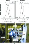
(a) Beam profiles of the microbeam BL32XU at SPring-8. A gold wire of 200 µm in diameter was used in the knife-edge scanning method (Mimura et al., 2007 ▸). The wire was scanned with translation axes with 10 nm positioning accuracy and X-ray signals were detected with a PIN photodiode. (b) Photograph of the experimental station of BL32XU.
Figure 4
Figure 4
Schematic drawing of data-collection strategies and each change of diffraction intensity from a single crystal using a microbeam. The total image number in single-point data collection is limited by radiation damage. Multi-point data collection can increase the total image number by avoiding serious radiation damage by using a fresh crystal volume with translation of the irradiation point. Helical data collection enables an equal distribution of radiation damage over the entire crystal volume. Thus, the helical method gives completely smoothed scales and B factors for frames, which finally improves the data quality.
Figure 5
Figure 5
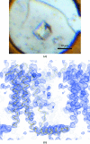
A single in meso microcrystal sufficient for structure solution by two-wavelength Hg-MAD at BL32XU. (a) A crystal of Hg-YidC. (b) The initial MAD-phased electron density at 1.0σ with a backbone of the final model of YidC. This was obtained by the helical data-collection method.
Figure 6
Figure 6
Automated microcrystal data-collection process. Firstly, the cryoloop is automatically centred and aligned under an optical microscope, followed by a low-dose X-ray raster scan in the defined area. The crystal positions are recognized by analyzing diffraction spots. A small-wedge diffraction dataset is collected from each crystal and processed to the merging of datasets to obtain complete and consistent data.
Figure 7
Figure 7
Various sample-delivery methods for serial synchrotron crystallography. (a) Liquid stream of a crystal suspension (Botha et al., 2015; Nogly et al., 2015 ▸), (b) loop-harvested cryocooled microcrystals (Gati et al., 2014; Hasegawa et al., 2017 ▸) and (c) thin-film substrate on which microcrystals are loaded (Coquelle et al., 2015 ▸).

References

    1. Assmann, G., Brehm, W. & Diederichs, K. (2016). J. Appl. Cryst. 49, 1021–1028. - PMC - PubMed
    1. Axford, D., Aller, P., Sanchez-Weatherby, J. & Sandy, J. (2016). Acta Cryst. F72, 313–319. - PMC - PubMed
    1. Axford, D., Foadi, J., Hu, N.-J., Choudhury, H. G., Iwata, S., Beis, K., Evans, G. & Alguel, Y. (2015). Acta Cryst. D71, 1228–1237. - PMC - PubMed
    1. Barty, A., Kirian, R. A., Maia, F. R. N. C., Hantke, M., Yoon, C. H., White, T. A. & Chapman, H. (2014). J. Appl. Cryst. 47, 1118–1131. - PMC - PubMed
    1. Batyuk, A. et al. (2016). Sci. Adv. 2, 2375–2548.