. 2017 Aug 31;4(Pt 5):678-694.
doi: 10.1107/S2052252517010922. eCollection 2017 Sep 1.

Single-particle cryo-EM using alignment by classification (ABC): the structure of Lumbricus terrestris haemoglobin

Affiliations

Single-particle cryo-EM using alignment by classification (ABC): the structure of Lumbricus terrestris haemoglobin

Pavel Afanasyev et al. IUCrJ. .

Abstract

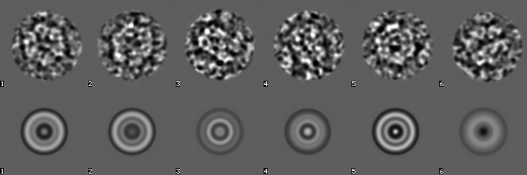
Single-particle cryogenic electron microscopy (cryo-EM) can now yield near-atomic resolution structures of biological complexes. However, the reference-based alignment algorithms commonly used in cryo-EM suffer from reference bias, limiting their applicability (also known as the 'Einstein from random noise' problem). Low-dose cryo-EM therefore requires robust and objective approaches to reveal the structural information contained in the extremely noisy data, especially when dealing with small structures. A reference-free pipeline is presented for obtaining near-atomic resolution three-dimensional reconstructions from heterogeneous ('four-dimensional') cryo-EM data sets. The methodologies integrated in this pipeline include a posteriori camera correction, movie-based full-data-set contrast transfer function determination, movie-alignment algorithms, (Fourier-space) multivariate statistical data compression and unsupervised classification, 'random-startup' three-dimensional reconstructions, four-dimensional structural refinements and Fourier shell correlation criteria for evaluating anisotropic resolution. The procedures exclusively use information emerging from the data set itself, without external 'starting models'. Euler-angle assignments are performed by angular reconstitution rather than by the inherently slower projection-matching approaches. The comprehensive 'ABC-4D' pipeline is based on the two-dimensional reference-free 'alignment by classification' (ABC) approach, where similar images in similar orientations are grouped by unsupervised classification. Some fundamental differences between X-ray crystallography versus single-particle cryo-EM data collection and data processing are discussed. The structure of the giant haemoglobin from Lumbricus terrestris at a global resolution of ∼3.8 Å is presented as an example of the use of the ABC-4D procedure.

Keywords: MSA; alignment by classification; angular reconstitution; cryo-EM; worm haemoglobin.


Figures

Figure 1
Figure 1
Full data set statistics. (a) Typical micrograph (the average of a seven-frame movie; coarsened to a size of 512 × 512) of the diluted worm-haemoglobin haemolymph. The images contain concentrated particles as well as some ‘junk’. Histograms of the average density (b) and the standard deviation (c) of all of the frames in the data set (36 645 frames or a total of 5235 movies). Images falling within the standard-deviation range marked in red were used for characterization of the camera properties.
Figure 2
Figure 2
Anisotropic magnification of the worm-haemoglobin data set became evident from an eigenvector analysis of all amplitude spectra (see §2.5). The second eigenimage revealed a 2.6% ellipticity of the ∼3.6 Å water ring (marked with large arrow in the left spectrum) and the 2.2 Å water ring (medium-sized arrow). A further water/ice ring can be seen at 1.8 Å (small arrow), which is only visible in the corners of the image (beyond the Nyquist frequency) but reflects back into the image everywhere else owing to aliasing (grey arrow). The right spectrum is the same as the left spectrum but is mirrored horizontally, thus changing the ellipticity direction. The ellipticity is in a close-to-diagonal (∼36°) direction such that the vertical and horizontal magnifications in the data set are almost identical. The Thon rings visible in the centre are elliptical owing to an astigmatism of ∼1000 Å. This astigmatism ellipticity is unrelated to the ellipticity of the water rings. The two effects may become entangled if the anisotropic magnification is not corrected prior to CTF determination.
Figure 3
Figure 3
(a) Selected class average of individual spectra with fitted theoretical spectrum. Top left, experimental class-average spectrum (1280 × 1280 patches). Bottom right, theoretical CTF amplitudes at a defocus of 7913/9047 Å (astigmatism of 1134 Å at an angle of 133°). Thon rings in good spectra are visible to beyond ∼2/3 of the Nyquist frequency, corresponding to a resolution of ∼3.1 Å. (b) Histogram of the defocus values of ∼450 000 movie-frame patches. The peak at the low-defocus edge of this histogram corresponds to outliers with a defocus less than 0.22 µm (the search limit), which were ignored.
Figure 4
Figure 4
Two spectra of drifted movies: (a) before and (b) after movie alignments. The second movie in (a) experienced a stronger drift, impeding the restoration of the high-resolution information (only two ‘zeroes’ in the direction of the drift were restored).
Figure 5
Figure 5
Six manually selected particles (top row) used as templates for an initial competitive particle picking and their rotational averages (bottom row).
Figure 6
Figure 6
The first 32 eigenimages of 20 000 picked particles. The first eigenimage is rotationally symmetric, reflecting this property of the first set of particle-picking templates. Eigenimages 2 and 3 reflect the predominant sixfold symmetry of the worm-haemoglobin ‘top views’.
Figure 7
Figure 7
15 class averages of unaligned particles (‘alignment by classification’; ABC) used for an initial ‘random-startup’ three-dimensional reconstruction. Class average 2 corresponds to a top view and class averages 3 and 10 correspond to side views. The other class averages represent various intermediate projection images of the haemoglobin. Since the input images are coarsened (binned) by a factor of four, no information beyond the Nyquist frequency of 1/8.8 Å can be present in the images at this stage.
Figure 8
Figure 8
Five class averages used for random startup (top row of Fig. 7 ▸) and the corresponding reprojections from the three-dimensional reconstruction. A good correspondence of the class averages to their reprojections is a necessary condition for a valid reconstruction procedure.
Figure 9
Figure 9
(a) Class averages randomly extracted from the total of 3000. High-contrast class averages are associated with good views of the worm haemoglobin, whereas low-contrast class averages are associated with atypical false-positive particle detections. (b) The histogram of standard deviations of 3000 class averages (205 764 particles) reveals a bimodal distribution in which virtually all remaining false positives land in low-contrast class averages.
Figure 10
Figure 10
Effect of ABC optimization. After ABC alignment of the full data set, all images are rotated and shifted to a common origin as dictated by the Euler orientations of the class averages. A new MSA classification thus yields a better class average since more molecular images now have a common three-dimensional orientation. Shown here are class averages at an intermediate level of the refinement procedures at a coarsening level of 2 (Nyquist frequency 1/4.4 Å).
Figure 11
Figure 11
FSC curves of the final three-dimensional reconstruction. (0.6 of the Nyquist frequency corresponds to 1/3.7 Å.) FSCβ<60° is based on the preferred top-view class averages (0 < β < 60°); FSCβ>60° assesses the contributions of the side-view class averages (60 < β < 90°). The circle indicates the area in which a high FSC value is a good indicator of the quality of the reconstruction.
Figure 12
Figure 12
Final cryo-EM map of worm haemoglobin (scale bars indicate 50 Å). (a) Slice through the cryo-EM three-dimensional reconstruction perpendicular to the main sixfold symmetry axis, with the yellow arrow pointing at a haem group sandwiched between two α-helices; the proximal histidine contacting the iron in the haem group is visible. (b) Surface rendering of the top view (along the sixfold axis); the asymmetric unit (1/12th of the D6 reconstruction; see Supplementary Movie S1) is marked in yellow. (c) The asymmetric unit (‘protomer’ or ‘1/12th unit’) with arrows pointing at the globin fold shown in (d) and the α-helix shown in (e). (d) View of one the better resolved haem groups: both the proximal and the distal histidine (b globin chain: His96 and His64) are well embedded in density. (e) View of one α-helix (L1 linker chain; Arg9–Leu38) in the triple coiled-coil region: the side chains fit nicely into the three-dimensional reconstruction. The atomic model was deposited as PDB entry 5m3l.
Figure 13
Figure 13
Fourier shell correlation plot of the model (protomer) versus the map (masked 1/12th subunit; van Heel et al., 2000 ▸). The atomic model was deposited as PDB entry 5m3l.

References

    1. Adams, P. D. et al. (2010). Acta Cryst. D66, 213–221. - PubMed
    1. Adrian, M., Dubochet, J., Lepault, J. & McDowall, A. W. (1984). Nature (London), 308, 32–36. - PubMed
    1. Afanasyev, P., Ravelli, R. B. G., Matadeen, R., De Carlo, S., van Duinen, G., Alewijnse, B., Peters, P. J., Abrahams, J.-P., Portugal, R. V., Schatz, M. & van Heel, M. (2015). Sci. Rep. 5, 10317. - PMC - PubMed
    1. Allegretti, M., Mills, D. J., McMullan, G., Kühlbrandt, W. & Vonck, J. (2014). Elife, 3, e01963. - PMC - PubMed
    1. Bai, X.-C., McMullan, G. & Scheres, S. H. W. (2015). Trends Biochem. Sci. 40, 49–57. - PubMed