

Blood pressure gradients in cerebral arteries: a clue to pathogenesis of cerebral small vessel disease
- PMID: 28989801
- PMCID: PMC5628379
- DOI: 10.1136/svn-2017-000087
Blood pressure gradients in cerebral arteries: a clue to pathogenesis of cerebral small vessel disease
Abstract
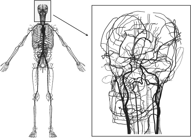
Rationale: The role of hypertension in cerebral small vessel disease is poorly understood. At the base of the brain (the 'vascular centrencephalon'), short straight arteries transmit blood pressure directly to small resistance vessels; the cerebral convexity is supplied by long arteries with many branches, resulting in a drop in blood pressure. Hypertensive small vessel disease (lipohyalinosis) causes the classically described lacunar infarctions at the base of the brain; however, periventricular white matter intensities (WMIs) seen on MRI and WMI in subcortical areas over the convexity, which are often also called 'lacunes', probably have different aetiologies.
Objectives: We studied pressure gradients from proximal to distal regions of the cerebral vasculature by mathematical modelling.
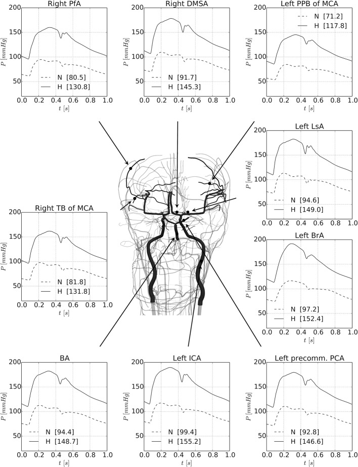
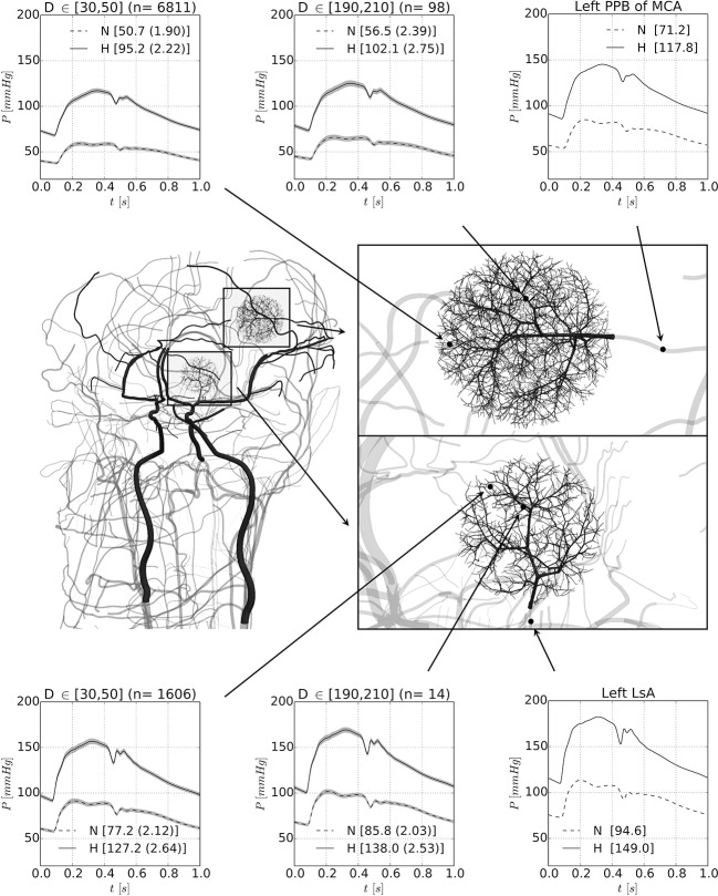
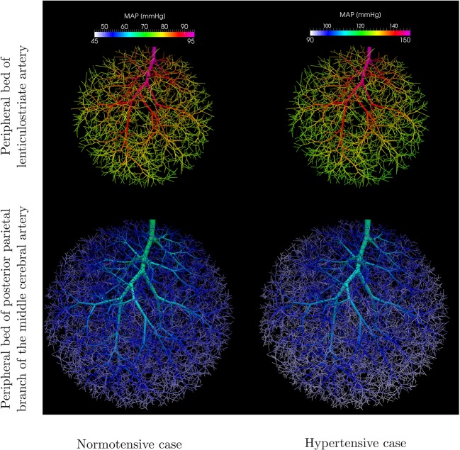
Methods and results: Blood flow/pressure equations were solved in an Anatomically Detailed Arterial Network (ADAN) model, considering a normotensive and a hypertensive case. Model parameters were suitably modified to account for structural changes in arterial vessels in the hypertensive scenario. Computations predict a marked drop in blood pressure from large and medium-sized cerebral vessels to cerebral peripheral beds. When blood pressure in the brachial artery is 192/113 mm Hg, the pressure in the small arterioles of the posterior parietal artery bed would be only 117/68 mm Hg. In the normotensive case, with blood pressure in the brachial artery of 117/75 mm Hg, the pressure in small parietal arterioles would be only 59/38 mm Hg.
Conclusion: These findings have important implications for understanding small vessel disease. The marked pressure gradient across cerebral arteries should be taken into account when evaluating the pathogenesis of small WMIs on MRI. Hypertensive small vessel disease, affecting the arterioles at the base of the brain should be distinguished from small vessel disease in subcortical regions of the convexity and venous disease in the periventricular white matter.
Keywords: Small vessel disease; amyloid; blood pressure; lacunar; lobar; mathematicalmodelling.
Conflict of interest statement
Competing interests: None declared.
Figures
References
-
- Fisher CM. Lacunar strokes and infarcts: a review. Neurology 1982;32:871–6. doi:10.1212/WNL.32.8.871 - DOI - PubMed
-
- Hachinski VC, Norris JW. The vascular infrastructure. The acute stroke. Philadelphia: F.A. Davis 1985. p. :27–40.
-
- Meier IB, Gu Y, Guzaman VA, et al. . Lobar microbleeds are associated with a decline in executive functioning in older adults. Cerebrovasc Dis 2014;38:377–83. doi:10.1159/000368998 - DOI - PMC - PubMed
-
- Caplan LR. Lacunar infarction and small vessel disease: pathology and pathophysiology. J Stroke 2015;17:2–6. doi:10.5853/jos.2015.17.1.2 - DOI - PMC - PubMed
-
- Lam TD, Lammers S, Munoz C, et al. . Diabetes, intracranial Stenosis and microemboli in asymptomatic carotid Stenosis. Can J Neurol Sci 2013;40:177–81. doi:10.1017/S031716710001369X - DOI - PubMed
MeSH terms
LinkOut - more resources
Full Text Sources
Other Literature Sources
Medical