

Volumetric accuracy of cone-beam computed tomography
- PMID: 28989899
- PMCID: PMC5620461
- DOI: 10.5624/isd.2017.47.3.165
Volumetric accuracy of cone-beam computed tomography
Abstract
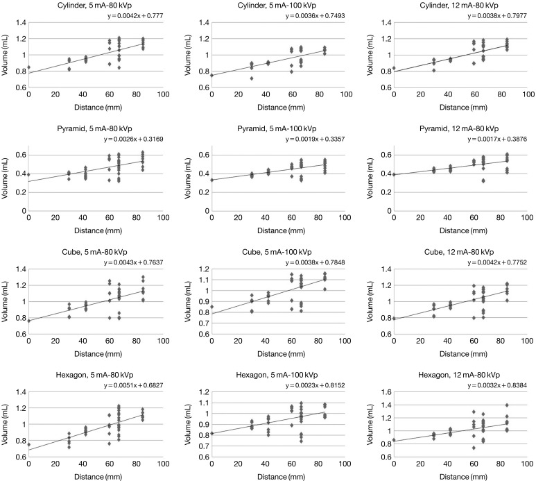
Purpose: This study was performed to investigate the influence of object shape and distance from the center of the image on the volumetric accuracy of cone-beam computed tomography (CBCT) scans, according to different parameters of tube voltage and current.
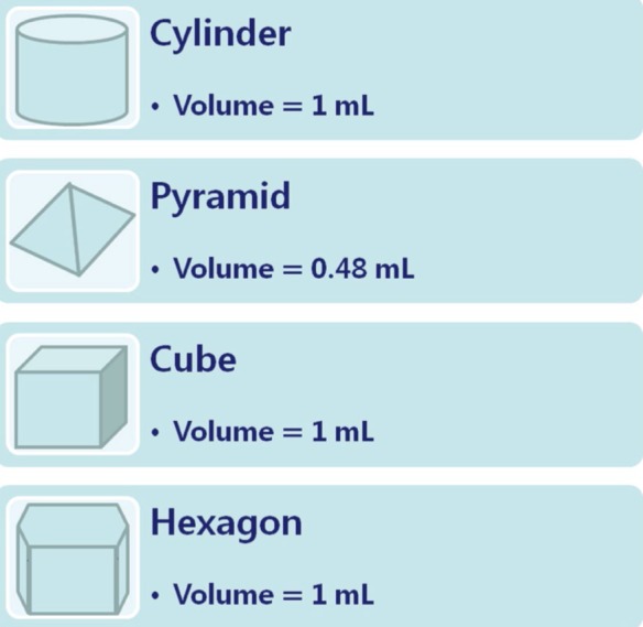
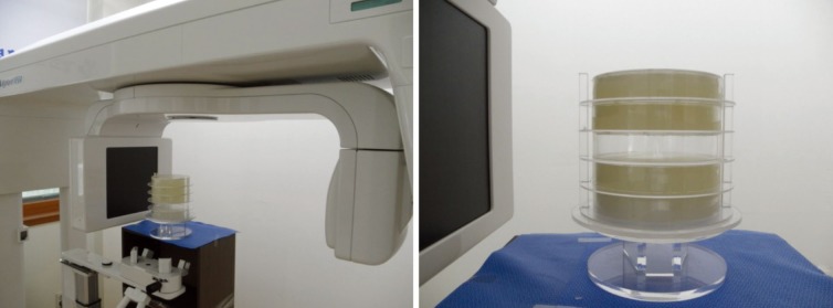
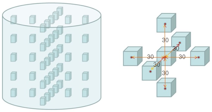
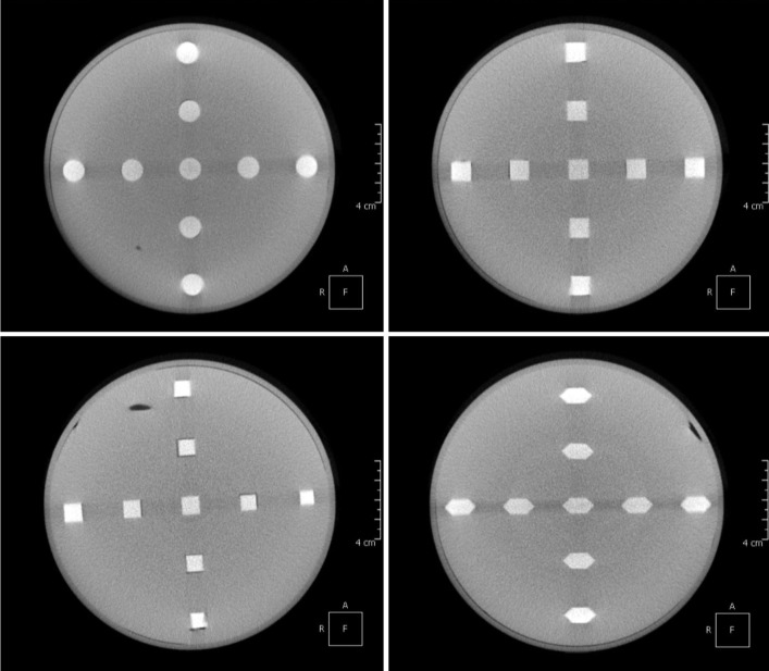
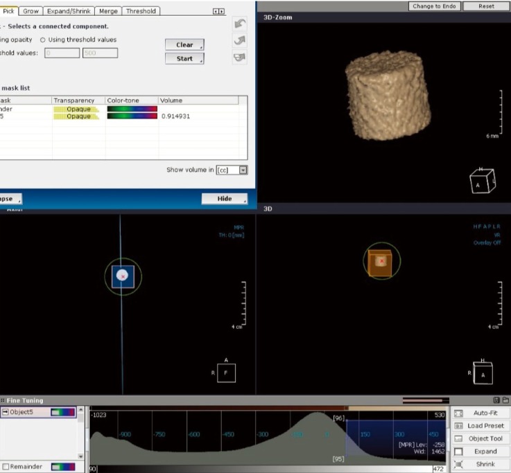
Materials and methods: Four geometric objects (cylinder, cube, pyramid, and hexagon) with predefined dimensions were fabricated. The objects consisted of Teflon-perfluoroalkoxy embedded in a hydrocolloid matrix (Dupli-Coe-Loid TM; GC America Inc., Alsip, IL, USA), encased in an acrylic resin cylinder assembly. An Alphard Vega Dental CT system (Asahi Roentgen Ind. Co., Ltd, Kyoto, Japan) was used to acquire CBCT images. OnDemand 3D (CyberMed Inc., Seoul, Korea) software was used for object segmentation and image analysis. The accuracy was expressed by the volume error (VE). The VE was calculated under 3 different exposure settings. The measured volumes of the objects were compared to the true volumes for statistical analysis.
Results: The mean VE ranged from -4.47% to 2.35%. There was no significant relationship between an object's shape and the VE. A significant correlation was found between the distance of the object to the center of the image and the VE. Tube voltage affected the volume measurements and the VE, but tube current did not.
Conclusion: The evaluated CBCT device provided satisfactory volume measurements. To assess volume measurements, it might be sufficient to use serial scans with a high resolution, but a low dose. This information may provide useful guidance for assessing volume measurements.
Keywords: Cone-Beam Computed Tomography; Imaging, Three-Dimensional; Phantom.
Figures
Similar articles
-
The incidence and configuration of the bifid mandibular canal in Koreans by using cone-beam computed tomography.Imaging Sci Dent. 2014 Mar;44(1):53-60. doi: 10.5624/isd.2014.44.1.53. Epub 2014 Mar 19. Imaging Sci Dent. 2014. PMID: 24701459 Free PMC article.
-
Evaluation of the cone beam CT for internal target volume localization in lung stereotactic radiotherapy in comparison with 4D MIP images.Med Phys. 2013 Nov;40(11):111709. doi: 10.1118/1.4823785. Med Phys. 2013. PMID: 24320417
-
Relationship between density variability and imaging volume size in cone-beam computerized tomographic scanning of the maxillofacial region: an in vitro study.Oral Surg Oral Med Oral Pathol Oral Radiol Endod. 2009 Mar;107(3):420-5. doi: 10.1016/j.tripleo.2008.05.049. Epub 2008 Aug 20. Oral Surg Oral Med Oral Pathol Oral Radiol Endod. 2009. PMID: 18715805
-
A new age estimation procedure based on the 3D CBCT study of the pulp cavity and hard tissues of the teeth for forensic purposes: A pilot study.J Forensic Leg Med. 2015 Nov;36:150-7. doi: 10.1016/j.jflm.2015.09.015. Epub 2015 Sep 30. J Forensic Leg Med. 2015. PMID: 26458182
-
Change in Image Quality According to the 3D Locations of a CBCT Phantom.PLoS One. 2016 Apr 19;11(4):e0153884. doi: 10.1371/journal.pone.0153884. eCollection 2016. PLoS One. 2016. PMID: 27093639 Free PMC article.
Cited by
-
Comparative Evaluation of Temporomandibular Joint Parameters in Unilateral and Bilateral Cleft Lip and Palate Patients Using Cone-Beam CT: Focus on Growing vs. Non-Growing Subjects.Healthcare (Basel). 2024 Aug 7;12(16):1563. doi: 10.3390/healthcare12161563. Healthcare (Basel). 2024. PMID: 39201121 Free PMC article.
-
Accuracy of upper airway volume measurements using different software products: a comparative analysis.Dentomaxillofac Radiol. 2025 Jul 1;54(5):350-356. doi: 10.1093/dmfr/twaf023. Dentomaxillofac Radiol. 2025. PMID: 40084997 Free PMC article.
-
A study on the association between the inferior nasal turbinate volume and the maxillary sinus mucosal lining using cone beam tomography.Heliyon. 2022 Mar 26;8(3):e09190. doi: 10.1016/j.heliyon.2022.e09190. eCollection 2022 Mar. Heliyon. 2022. PMID: 35368549 Free PMC article.
-
AI-based automatic segmentation of craniomaxillofacial anatomy from CBCT scans for automatic detection of pharyngeal airway evaluations in OSA patients.Sci Rep. 2022 Jul 13;12(1):11863. doi: 10.1038/s41598-022-15920-1. Sci Rep. 2022. PMID: 35831451 Free PMC article.
-
Associations between the Volume of Bilateral Inferior Turbinates with Ipsilateral and Contralateral Sinus Mucosal Lining Thicknesses in Various Ages and Sexes: a CBCT Study of 302 Individuals.Maedica (Bucur). 2024 Sep;19(3):551-560. doi: 10.26574/maedica.2024.19.3.551. Maedica (Bucur). 2024. PMID: 39553361 Free PMC article.
References
-
- Palomo L, Palomo JM. Cone beam CT for diagnosis and treatment planning in trauma cases. Dent Clin North Am. 2009;53:717–727. - PubMed
-
- Scarfe WC, Farman AG, Sukovic P. Clinical applications of cone-beam computed tomography in dental practice. J Can Dent Assoc. 2006;72:75–80. - PubMed
-
- Landi A, Marina R, DeGrandi C, Crespi A, Montanari G, Sganzerla EP, et al. Accuracy of stereotactic localization with magnetic resonance compared to CT scan: experimental findings. Acta Neurochir (Wien) 2001;143:593–601. - PubMed
-
- Karger CP, Hipp P, Henze M, Echner G, Hoss A, Schad L, et al. Stereotactic imaging for radiotherapy: accuracy of CT, MRI, PET and SPECT. Phys Med Biol. 2003;48:211–221. - PubMed
-
- Hilgers ML, Scarfe WC, Scheetz JP, Farman AG. Accuracy of linear temporomandibular joint measurements with cone beam computed tomography and digital cephalometric radiography. Am J Orthod Dentofacial Orthop. 2005;128:803–811. - PubMed
LinkOut - more resources
Full Text Sources
Other Literature Sources
Miscellaneous
