

Analysis of the root position of the maxillary incisors in the alveolar bone using cone-beam computed tomography
- PMID: 28989901
- PMCID: PMC5620463
- DOI: 10.5624/isd.2017.47.3.181
Analysis of the root position of the maxillary incisors in the alveolar bone using cone-beam computed tomography
Abstract
Purpose: The purpose of this study was to measure the buccal bone thickness and angulation of the maxillary incisors and to analyze the correlation between these parameters and the root position in the alveolar bone using cone-beam computed tomography (CBCT).
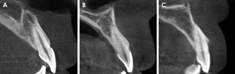
Materials and methods: CBCT images of 398 maxillary central and lateral incisors from 199 patients were retrospectively reviewed. The root position in the alveolar bone was classified as buccal, middle, or palatal, and the buccal type was further classified into subtypes I, II, and III. In addition, the buccolingual inclination of the tooth and buccal bone thickness were evaluated.
Results: A majority of the maxillary incisors were positioned more buccally within the alveolar bone, and only 2 lateral incisors (0.5%) were positioned more palatally. The angulation of buccal subtype III was the greatest and that of the middle type was the lowest. Most of the maxillary incisors exhibited a thin facial bone wall, and the lateral incisors had a significantly thinner buccal bone than the central incisors. The buccal bone of buccal subtypes II and III was significantly thinner than that of buccal subtype I.
Conclusion: A majority of the maxillary incisor roots were positioned close to the buccal cortical plate and had a thin buccal bone wall. Significant relationships were observed between the root position in the alveolar bone, the angulation of the tooth in the alveolar bone, and buccal bone thickness. CBCT analyses of the buccal bone and sagittal root position are recommended for the selection of the appropriate treatment approach.
Keywords: Cone-Beam Computed Tomography; Incisor; Maxilla; Tooth Root.
Figures


References
-
- Chan HL, Garaicoa-Pazmino C, Suarez F, Monje A, Benavides E, Oh TJ, et al. Incidence of implant buccal plate fenestration in the esthetic zone: a cone beam computed tomography study. Int J Oral Maxillofac Implants. 2014;29:171–177. - PubMed
-
- Lau SL, Chow J, Li W, Chow LK. Classification of maxillary central incisors-implications for immediate implant in the esthetic zone. J Oral Maxillofac Surg. 2011;69:142–153. - PubMed
-
- Wang HM, Shen JW, Yu MF, Chen XY, Jiang QH, He FM. Analysis of facial bone wall dimensions and sagittal root position in the maxillary esthetic zone: a retrospective study using cone beam computed tomography. Int J Oral Maxillofac Implants. 2014;29:1123–1129. - PubMed
-
- Braut V, Bornstein MM, Belser U, Buser D. Thickness of the anterior maxillary facial bone wall-a retrospective radiographic study using cone beam computed tomography. Int J Periodontics Restorative Dent. 2011;31:125–131. - PubMed
-
- Kan JY, Roe P, Rungcharassaeng K, Patel RD, Waki T, Lozada JL, et al. Classification of sagittal root position in relation to the anterior maxillary osseous housing for immediate implant placement: a cone beam computed tomography study. Int J Oral Maxillofac Implants. 2011;26:873–876. - PubMed
LinkOut - more resources
Full Text Sources
Other Literature Sources
