Evaluation of Progesterone and Ovulation-stimulating Drugs on the Glandular Epithelium and Angiogenesis in Mice
- PMID: 28989909
- PMCID: PMC5627566
- DOI: 10.4103/abr.abr_179_16
Evaluation of Progesterone and Ovulation-stimulating Drugs on the Glandular Epithelium and Angiogenesis in Mice
Abstract
Background: Human endometrium is a dynamic tissue during the menstrual cycle can be influenced by ovarian hormones. The purpose of this study was to evaluate the endometrium angiogenesis under the influence of human menopausal gonadotropin and human chorionic gonadotropin (HMG and HCG) that stimulate ovulation and progesterone.
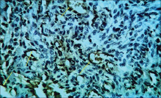
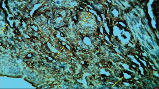
Materials and methods: In this study, thirty adult female mice were randomly divided into three groups as: control, gonadotropin and gonadotropin + progesterone. The mice in the other two groups except the control group received 7.5 IU HMG and later HCG. Subsequently, the mice were placed in a cage for mating. Gonadotropin + progesterone group was administered, 1 mg/mouse progesterone in 24, 48, and 72 h interval, after HMG injection. Ninety-six hours after HMG injection, animals were sacrificed, and their uterine specimens were prepared by immunohistochemistry technique for light microscopic studies, and statistical analysis was carried out.
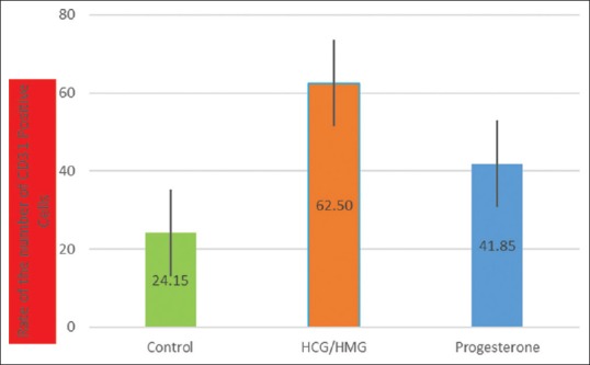
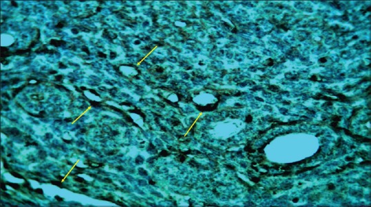
Results: Endometrium angiogenesis in control group showed that mean ± standard deviation was 24.15 ± 11.15, gonadotropin group was 62.50 ± 24.16, and gonadotropin + progesterone group was 41.85 ± 19.54. Significant difference between the control group and gonadotropin group and between the control group and gonadotropin + progesterone was observed. Statistically significant differences were observed in all groups in the endometrial angiogenesis (P < 0.05).
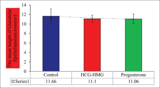
Conclusion: Ovarian induction with gonadotropins and gonadotropins + progesterone could not change the morphometrically index of endometrial glandular epithelium in mice. Ovarian stimulation followed by progesterone injection could modify the angiogenesis of mice endometrium.
Keywords: Angiogenesis; endometrium; implantation; progesterone.
Conflict of interest statement
There are no conflicts of interest.
Figures
References
-
- Martínez-Conejero JA, Simón C, Pellicer A, Horcajadas JA. Is ovarian stimulation detrimental to the endometrium? Reprod Biomed Online. 2007;15:45–50. - PubMed
-
- Wang H, Dey SK. Roadmap to embryo implantation: Clues from mouse models. Nat Rev Genet. 2006;7:185–99. - PubMed
-
- Nikas G, Makrigiannakis A. Endometrial pinopodes and uterine receptivity. Ann N Y Acad Sci. 2003;997:120–3. - PubMed
-
- Mulac-Jericevic B, Conneely OM. Reproductive tissue selective actions of progesterone receptors. Reproduction. 2004;128:139–46. - PubMed
-
- Mercé LT, Barco MJ, Bau S, Troyano J. Are endometrial parameters by three-dimensional ultrasound and power Doppler angiography related to in vitro fertilization/embryo transfer outcome? Fertil Steril. 2008;89:111–7. - PubMed
LinkOut - more resources
Full Text Sources
Other Literature Sources