

Liver lesions detected in a hepatitis B core total antibody-positive patient masquerading as hepatocellular carcinoma: a rare case of peliosis hepatis and a review of the literature
- PMID: 28990003
- PMCID: PMC5620477
- DOI: 10.14701/ahbps.2017.21.3.157
Liver lesions detected in a hepatitis B core total antibody-positive patient masquerading as hepatocellular carcinoma: a rare case of peliosis hepatis and a review of the literature
Abstract
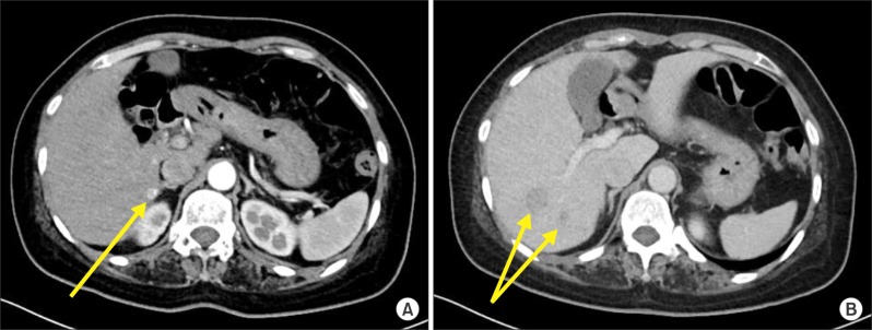
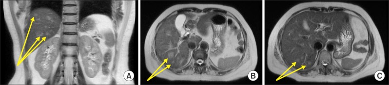
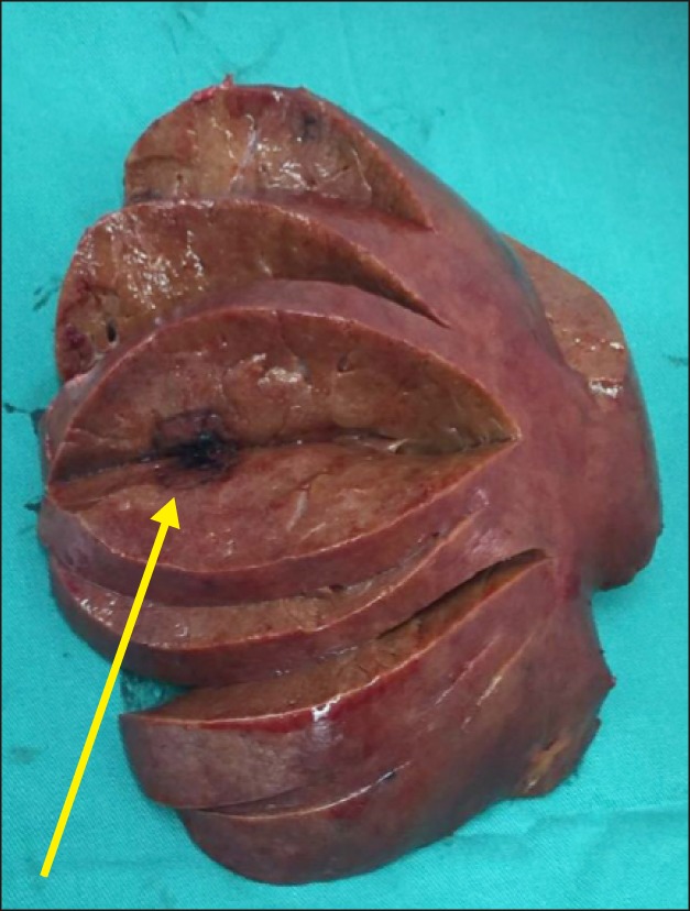
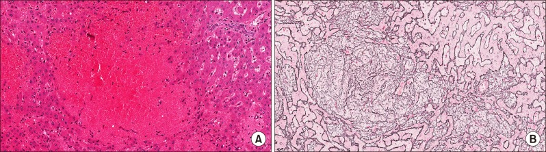
Peliosis Hepatis (PH) is a rare vascular disorder of the liver, characterized by the presence of cystic blood-filled cavities distributed throughout the hepatic parenchyma. The pathogenesis of PH remains controversial. The preoperative diagnosis of PH is difficult, due to the non-specific imaging characteristics of PH and almost all cases are diagnosed on histology post resection. This study presents a case of PH masquerading as hepatocellular carcinoma (HCC). The patient is a 45-year old Chinese lady, who presented with transaminitis. She was found to be hepatitis B virus core total antibody-positive with an alpha-fetoprotein (AFP) of 29.4 ng/ml. Triphasic liver computed tomography showed several arterial hypervascular lesions and hypoenhancing lesions on the venous phase, particularly in the segments 6/7. Subsequently, a magnetic resonance imaging scan showed multiple lesions in the right hemiliver with an indeterminate enhancement patterns. Subsequently, she decided to undergo a resection procedure. Histopathology revealed findings consistent with PH with some unusual features. This case demonstrates a clinical conundrum, in which PH presented with a raised AFP, in a patient with risk factors for the development of HCC. The clinical suspicion of PH should be high in patients, who present with multiple hepatic lesions with variable enhancement patterns.
Keywords: Hepatis; Hepatocellular carcinoma; Hepatoma; Peliosis.
Figures
References
Publication types
LinkOut - more resources
Full Text Sources
Other Literature Sources