. 2017 Dec;16(6):8868-8874.
doi: 10.3892/mmr.2017.7691. Epub 2017 Oct 3.

Mechanism of the effect of saikosaponin on atherosclerosis in vitro is based on the MAPK signaling pathway

Affiliations

Mechanism of the effect of saikosaponin on atherosclerosis in vitro is based on the MAPK signaling pathway

Lin Yang et al. Mol Med Rep. 2017 Dec.

Abstract

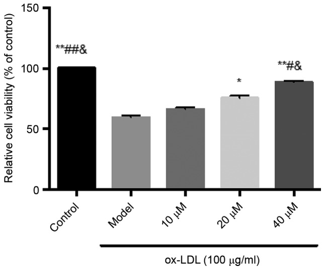
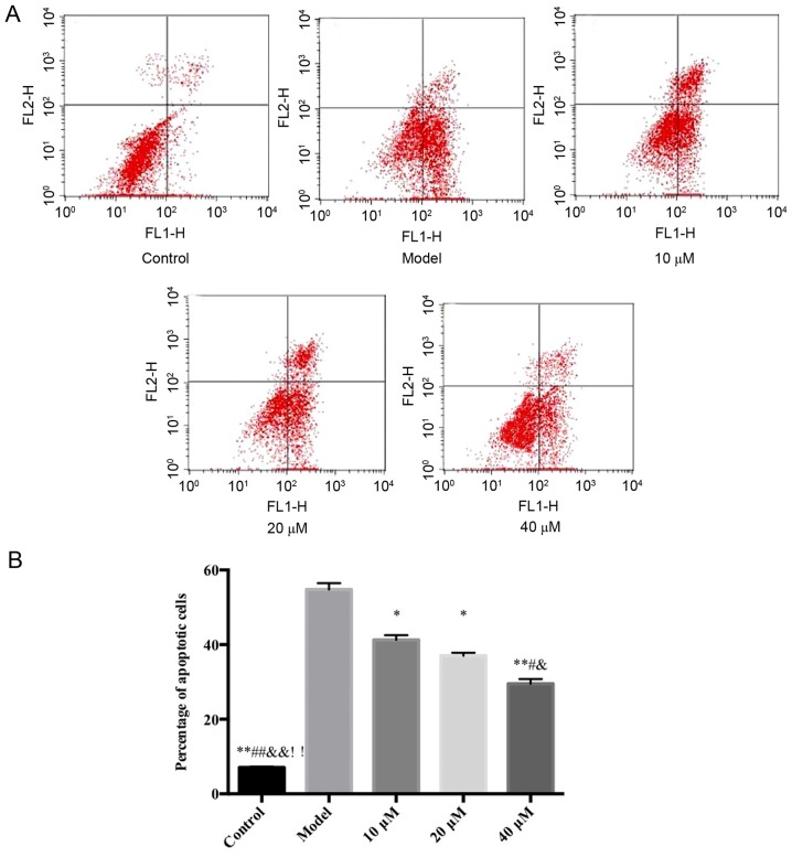
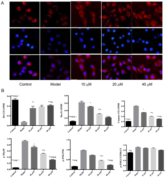
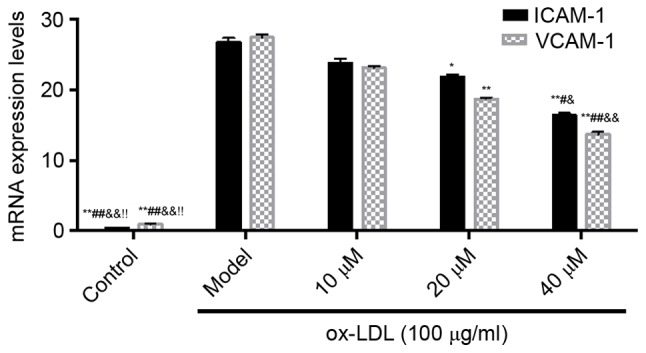
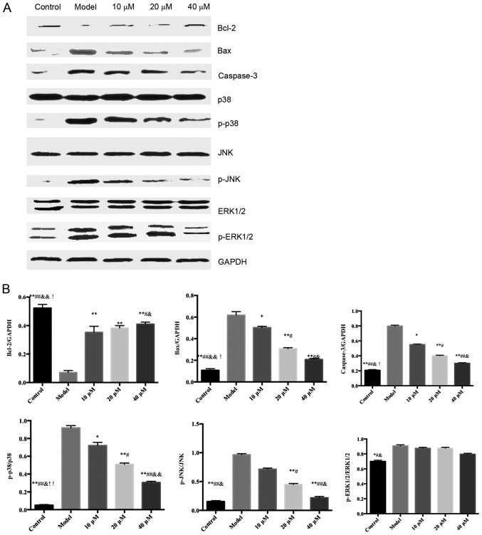
The present study aimed to investigate the effects of saikosaponin on oxidized low‑density lipoprotein (ox‑LDL)‑induced human umbilical vein endothelial cell (HUVEC) injury and apoptosis, and examine the involvement of the mitogen‑activated protein kinase (MAPK) signaling pathway. The viability and apoptosis of HUVECs were detected using an MTT assay and flow cytometry. ELISA analysis was applied to measure the levels of tumor necrosis factor (TNF)‑α and interleukin (IL)‑6 cytokines. Nuclear factor (NF)‑κB p65 nuclear translocation was observed using immunofluorescence staining. The levels of intercellular adhesion molecule 1 and vascular cell adhesion molecule‑1 were detected using reverse transcription‑polymerase chain reaction analysis. The phosphorylation of B‑cell lymphoma 2 (Bcl‑2), Bcl‑2‑associated X protein (Bax), caspase‑3 p38, c‑Jun N‑terminal kinase (JNK) and extracellular signal‑regulated kinase (ERK)1/2 were detected using western blot analysis. The results revealed that saikosaponin increased the viability of the HUVECs and decreased the early‑stage apoptotic rate of the HUVECs induced by ox‑LDL. The expression levels of inflammatory cytokines in the injured vascular endothelial cells were decreased, the expression levels of adhesion molecules were reduced, the activity of superoxide dismutase was increased, and malondialdehyde content was decreased. Therefore, the inflammatory response and oxidative stress were inhibited. Simultaneously, the levels of Bcl‑2 increased, the levels of Bax and caspase‑3 decreased, and the nuclear translocation of NF‑κB p65 was significantly inhibited. The protein levels of phosphorylated p38 and JNK were reduced, whereas that of ERK1/2 remained unaffected. It was concluded that the MAPK signaling pathway mediated HUVEC injury induced by ox‑LDL. However, saikosaponin inhibited the HUVEC injury induced by ox‑LDL through inhibiting the ERK1/2 and p38 MAPK signaling pathways, and possibly also through the JNK and p38 MAPK signaling pathway.


Figures

Figure 1.
Figure 1.
Inhibitory effects of saikosaponin on ox-LDL-induced human umbilical vein endothelial cell injury. **P<0.01 vs. Model; *P<0.05 vs. Model; ##P<0.01 vs. 10 µM; #P<0.05, vs. 10 µM; &P<0.05, vs. 20 µM. ox-LDL, oxidized low-density lipoprotein.
Figure 2.
Figure 2.
Flow cytometric analysis of the inhibitory effects of saikosaponin on oxidized low-density lipoprotein-induced human umbilical vein endothelial cell injury. (A) Flow cytometry data of control, model and 10, 20 and 40 µM saikosaponin treated human umbilical vein endothelial cells. (B) Quantitative results of flow cytometry data. ox-LDL, oxidized low density lipoprotein. *P<0.05 vs. model, **P<0.01 vs. model; #P<0.05 vs. 10 µM; ##P<0.01 vs. 10 µM; &P<0.05 vs. 20 µM; &&P<0.01 vs. 20 µM; !!P<0.01 vs. 40 µM.
Figure 3.
Figure 3.
Effect of saikosaponin on nuclear factor-κB p65 nuclear transfer. Magnification, ×200. (A) Immunofluorescence data of control, model and 10, 20 and 40 µM saikosaponin treated human umbilical vein endothelial cells. (B) Quantitative results of immunnofluorescence data. *P<0.05 vs. model, **P, 0.01 vs. model; #P<0.05 vs. 10 µM; ##P<0.01 vs. 10 µM; &P<0.05 vs. 20 µM; &&P<0.01 vs. 20 µM; !P<0.05 vs. 10 µM; !!P<0.01 vs. 40 µM.
Figure 4.
Figure 4.
Effects of saikosaponin on the mRNA expression levels of ICAM-1 and VCAM-1. **P<0.01, vs. Model; *P<0.05, vs. Model; ##P<0.01 vs. 10 µM; #P<0.05, vs. 10 µM; &&P<0.01 vs. 20 µM; &P<0.05 vs. 20 µM; !P<0.05 vs. 40 µM. ICAM-1, intercellular adhesion molecule 1; VCAM-1, vascular cell adhesion molecule-1; ox-LDL, oxidized low-density lipoprotein.
Figure 5.
Figure 5.
Effects of saikosaponin on the protein expression levels of caspase-3, Bcl-2, Bax, ERK1/2, p-ERK1/2, p38, p-p38, JNK and p-JNK. (A) Blots demonstrate the expression levels of proteins. (B) Quantitative results of the western blotting. *P<0.05 vs. model, **P<0.01 vs. model; #P<0.05 vs. 10 µM; ##P<0.01 vs. 10 µM; &P<0.05 vs. 20 µM; &&P<0.01 vs. 20 µM; !P<0.05 vs. 40 µM, !!P<0.01 vs. 40 µM. Bcl-2, B-cell lymphoma 2; Bax, Bcl-2-associated X protein; ERK, extracellular signal-regulated kinase; p-, phosphorylated.

Similar articles

Cited by

References

    1. Lee SM, Lee YJ, Kim YC, Kim JS, Kang DG, Lee HS. Vascular protective role of vitexicarpin isolated from Vitex rotundifolia in human umbilical vein endothelial cells. Inflammation. 2012;35:584–593. doi: 10.1007/s10753-011-9349-x. - DOI - PubMed
    1. Madonna R, Massaro M, Pandolfi A, Consoli A, De Caterina R. The prominent role of p38 mitogen-activated protein kinase in insulin-mediated enhancement of VCAM-1 expression in endothelial cells. Int J Immunopathol Pharmacol. 2007;20:539–555. doi: 10.1177/039463200702000312. - DOI - PubMed
    1. Wang Z, Niu Q, Peng X, Li M, Liu Y, Liu J, Wen S, Wei Y. Mitofusin 2 ameliorates aortic remodeling by suppressing ras/raf/ERK pathway and regulating mitochondrial function in vascular smooth muscle cells. Int J Cardiol. 2015;178:165–167. doi: 10.1016/j.ijcard.2014.10.122. - DOI - PubMed
    1. Park B, Yim JH, Lee HK, Kim BO, Pyo S. Ramalin inhibits VCAM-1 expression and adhesion of monocyte to vascular smooth muscle cells through MAPK and PADI4-dependent NF-κB and AP-1 pathways. Biosci Biotechnol Biochem. 2015;79:539–552. doi: 10.1080/09168451.2014.991681. - DOI - PubMed
    1. Martin-Blanco E. p38 MAPK signalling cascades: Ancient roles and new functions. Bioessays. 2000;22:637–645. doi: 10.1002/1521-1878(200007)22:7<637::AID-BIES6>3.0.CO;2-E. - DOI - PubMed

MeSH terms