. 2017 Dec;16(6):8930-8936.
doi: 10.3892/mmr.2017.7694. Epub 2017 Oct 3.

Kinin B1 receptor as a novel, prognostic progression biomarker for carotid atherosclerotic plaques

Affiliations

Kinin B1 receptor as a novel, prognostic progression biomarker for carotid atherosclerotic plaques

Tiantian Liu et al. Mol Med Rep. 2017 Dec.

Abstract

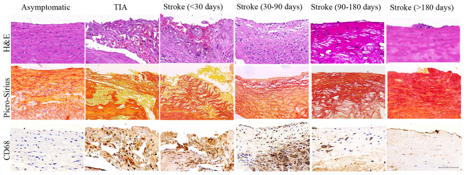
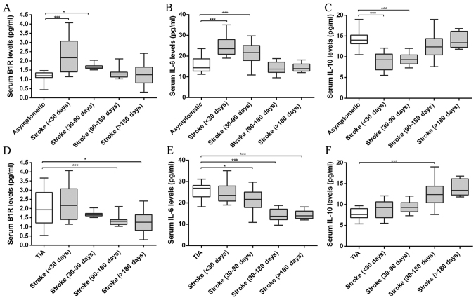
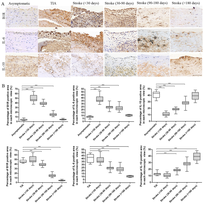
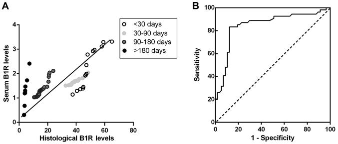
Stroke caused by atherosclerosis remains a leading cause of morbidity and mortality worldwide, associated with carotid plaque rupture and inflammation progression. However, the inflammatory biomarkers which aid in predicting the future course of plaques are less detailed. The present study investigated the association between plaque vulnerable and inflammatory biomarkers using blood and plaque specimens. Carotid plaque specimens were obtained from 80 patients following stroke, 14 patients suffering from transient ischaemic attack and 17 asymptomatic patients that underwent carotid endarterectomy. To assess changes in plaque characteristics at histological levels, plaques were categorized by the time between the latest ischemic stroke and surgical intervention within 30, 30‑90, 90‑180 and over 180 days following stroke. Serum levels of inflammatory biomarkers interleukin (IL)‑6, IL‑10 and kinin B1 receptor (B1R) were measured by ELISA. Histological assessment of plaque was used to evaluate the plaque stability, progression and the inflammatory biomarker levels. Comparisons of histological characteristics demonstrated that plaques revealed an unstable phenotype following stroke within 30, 30‑90 days and then remodeled into more stable plaques following stroke at 90‑180 and over 180 days. By comparing the serum levels of inflammatory biomarkers, it was observed that IL‑6 and B1R levels tended to decline whereas IL‑10 levels increased in stroke patients from <30 days to over 180 days. Immunohistochemical analysis of IL‑6, IL‑10 and B1R demonstrated similar alterations in serum levels. Correlation analyses revealed that only B1R serum level was significantly correlated with histological level in patients with carotid atherosclerosis. The findings revealed that serum B1R levels may provide prognostic information and currently act as potential indicators for progression in atherosclerosis.


Figures

Figure 1.
Figure 1.
Histological characteristics of plaques. Representative images of plaques in asymptomatic, TIA, stroke patients of <30, 30–90, 90–180 and >180 days. H&E staining showed inflammatory cell infiltrations (TIA, Stroke <30 d and Stroke 30–90 d groups) and intraplaque hemorrhage (TIA, Stroke <30 d groups) in the vulnerable plaques and intact cap and predominantly fibrous tissue in the stable plaques (Asymptomatic, Stroke 90–180 d and Stroke >180 d groups). Picro-Sirius Red staining showed the large cholesterol crystals in the lipid core in vulnerable plaques (TIA, Stroke <30 d and Stroke 30–90 d groups) and collagen fibrous tissue in stable plaques (Asymptomatic, Stroke 90–180 d and Stroke >180 d groups). CD 68 staining showed histological appearance of a plaque with substantial macrophage infiltration in the vulnerable plaques (TIA, Stroke <30 d and Stroke 30–90 d groups) and minor macrophage infiltration in stable plaques (Asymptomatic, Stroke 90–180 d and Stroke >180 d groups). Scale bar: 100 µm.
Figure 2.
Figure 2.
Serum levels of proinflammatory and anti-inflammatory markers in asymptomatic, TIA and stroke patients of <30, 30–90, 90–180 and >180 days measured by ELISA. (A and D) B1R levels of plaques in stroke patients of <30, 30–90, 90–180 and >180 days compared with asymptomatic or TIA patients. (B and E) IL-6 levels of plaques in stroke patients compared with asymptomatic or TIA patients. (C and F) IL-10 levels of plaques in stroke patients compared with asymptomatic or TIA patients. Compared with asymptomatic group, stroke patients showed increased B1R and IL-6 levels at <30 days and this increased serum B1R and IL-6 levels tended to decline after stroke from 30–90 to >180 days. By contrast, the anti- inflammatory marker IL-10 levels were gradually upregulated in stroke patients in spatiotemporal course from<30 days to >180 days. (*P<0.05, ***P<0.001 vs. asymptomatic or TIA group).
Figure 3.
Figure 3.
Immunohistochemical results. (A) Representative immunohistochemical images of B1R, IL-6 and IL-10 in plaques of asymptomatic, TIA and stroke patients of <30, 30–90, 90–180 and >180 days. (B) Quantification of immunohistochemistry analysis of B1R, IL-6 and IL-10 staining of section from asymptomatic, TIA and stroke patients of <30, 30–90, 90–180 and >180 days. Percentage of positive staining area in each microscopic view (at 200x) showed that stroke plaques experienced initial increases of B1R and IL-6 contents at <30 days, and subsequent reduction from 30–90 to >180 days. However, a noticeable increase in IL-10 contents was found in stroke plaques from <30 to >180 days. (***P<0.001 vs. asymptomatic or TIA group). Scale bar: 100 µm.
Figure 4.
Figure 4.
Serum B1R levels for predicting vulnerable plaque in patients of stroke groups. (A) Histological levels of B1R in individual patients were correlated with the serum values of B1R in Stroke groups of <30, 30–90, 90–180 and >180 days (r=0.54, P<0.001). The regression curve line and significance values are shown in the plots. (B) Receiver-operating curve of B1R level for predicting vulnerable plaque in patients. The area under the curve of B1R levels were obtained from stroke patients.

Similar articles

Cited by

References

    1. Rothstein L, Jickling GC. Ischemic stroke biomarkers in blood. Biomark Med. 2013;7:37–47. doi: 10.2217/bmm.12.104. - DOI - PubMed
    1. Mauriello A, Servadei F, Sangiorgi G, Anemona L, Giacobbi E, Liotti D, Spagnoli LG. Asymptomatic carotid plaque rupture with unexpected thrombosis over a non-canonical vulnerable lesion. Atherosclerosis. 2011;218:356–362. doi: 10.1016/j.atherosclerosis.2011.06.056. - DOI - PubMed
    1. Stefanadis C, Antoniou CK, Tsiachris D, Pietri P. Coronary atherosclerotic vulnerable plaque: Current perspectives. J Am Heart Assoc. 2017;6:pii: e005543. doi: 10.1161/JAHA.117.005543. - DOI - PMC - PubMed
    1. Tuttolomondo A, Pecoraro R, Casuccio A, Di Raimondo D, Buttà C, Clemente G, Corte V Della, Guggino G, Arnao V, Maida C, et al. Peripheral frequency of CD4+ CD28- cells in acute ischemic stroke: Relationship with stroke subtype and severity markers. Medicine (Baltimore) 2015;94:e813. doi: 10.1097/MD.0000000000000813. - DOI - PMC - PubMed
    1. Tuttolomondo A, Pedone C, Pinto A, Di Raimondo D, Fernandez P, Di Sciacca R, Licata G. Gruppo Italiano di Farmacoepidemiologia dell'Anziano (GIFA) researchers: Predictors of outcome in acute ischemic cerebrovascular syndromes: The GIFA study. Int J Cardiol. 2008;125:391–396. doi: 10.1016/j.ijcard.2007.03.109. - DOI - PubMed