. 2018 Jan;149(1):31-42.
doi: 10.1007/s00418-017-1608-y. Epub 2017 Oct 9.

Expression of matrix metalloproteinase 12 is highly specific for non-proliferating invasive trophoblasts in the first trimester and temporally regulated by oxygen-dependent mechanisms including HIF-1A

Affiliations

Expression of matrix metalloproteinase 12 is highly specific for non-proliferating invasive trophoblasts in the first trimester and temporally regulated by oxygen-dependent mechanisms including HIF-1A

Ursula Hiden et al. Histochem Cell Biol. 2018 Jan.

Erratum in

Abstract

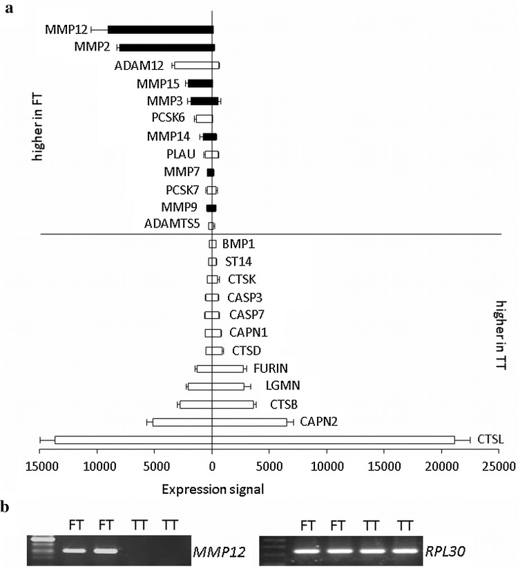
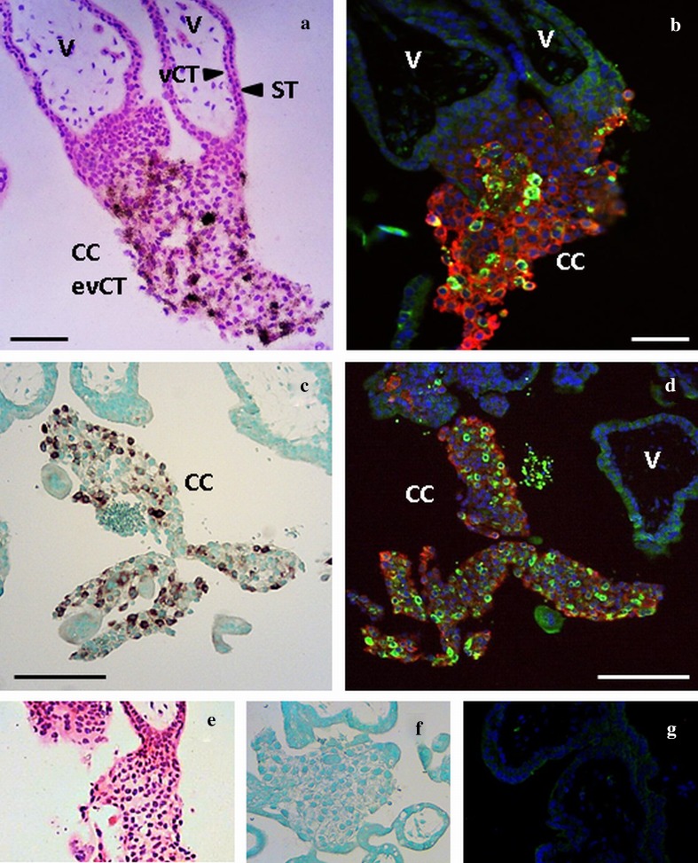
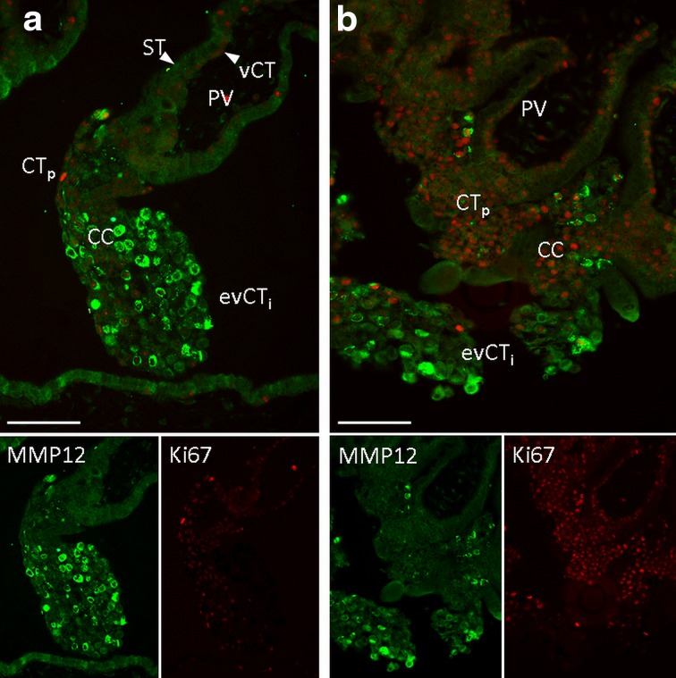
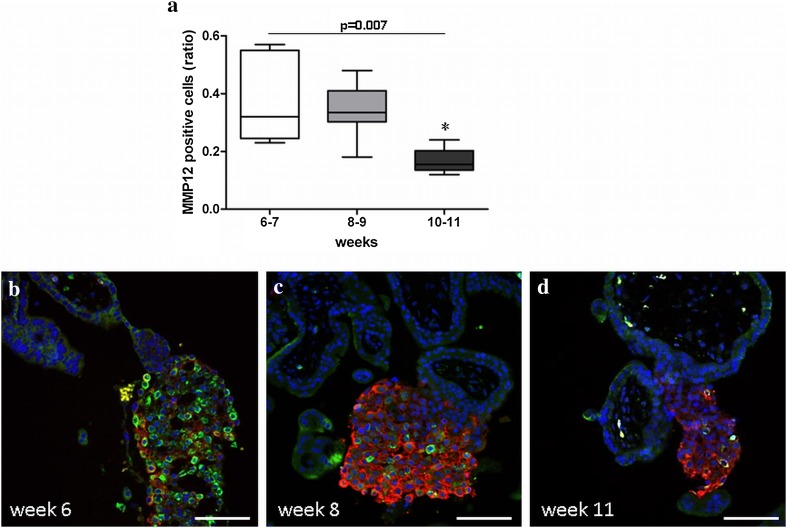
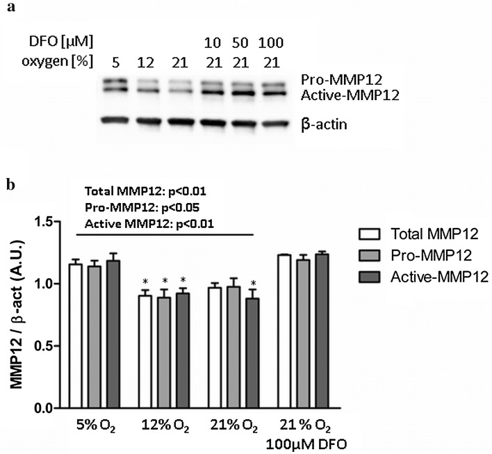
During first trimester pregnancy, trophoblast cells invade from the placenta into the maternal decidua where they anchor the placenta and remodel luminal structures like spiral arteries. This process depends on proteases secreted by invading trophoblasts, which degrade extracellular matrix (ECM). We here aimed to identify proteases particularly important for trophoblast invasion. We generated a list of proteases capable of degrading decidual ECM and trophoblast integrins using MEROPS database and compared expression of these proteases between primary trophoblasts isolated from first trimester placenta (FT, n = 3), representing an invasive phenotype, vs trophoblasts isolated from term pregnancy (TT, n = 3), representing a non-invasive trophoblast phenotype. Matrix metalloproteinase 12 (MMP12) revealed highest expression levels in FT, with absent expression in TT. In situ hybridisation and immunofluorescence localised MMP12 specifically to extravillous trophoblasts (evCT) whilst Ki67 co-staining revealed that proliferating trophoblasts of the cell columns were almost negative for MMP12. Quantification revealed a decline in MMP12 positive evCT at the end of first trimester, when oxygen levels start rising. MMP12 promoter analysis identified potential binding sites for hypoxia-inducible factor (HIF-1) and other oxygen-sensitive transcription factors. Moreover, MMP12 protein was increased by low oxygen in FT in vitro and by addition of a HIF-1α activator. Collectively, MMP12 is a highly expressed protease specific for invasive evCT during the first trimester. MMP12 down regulation by increasing oxygen concentration enables temporal expression control of MMP12 and involves several mechanisms including HIF-1α. These findings suggest MMP12 involved in trophoblast invasion during the first trimester.

Keywords: HIF-1α; Hypoxia; Matrix metalloproteinase 12; Trophoblast invasion.


Conflict of interest statement

The authors declare they have no conflict of interest.

Figures

Fig. 1
Fig. 1
(a) Proteases capable to degrade decidual ECM and integrins, expressed by primary trophoblasts from first trimester (FT) and term of gestation (TT). Among all proteases degrading collagens I, III, IV, V and VI, laminin, fibronectin, fibrillin 1 and 2, vitronectin and integrins specific for extravillous trophoblasts, MMP12 was highest expressed in FT with almost absent expression in TT. MMPs are coloured in black. Data are shown as mean ± SD. b The expression difference of MMP12 in primary first trimester (FT) vs. term trophoblasts (TT) was confirmed by semiquantitative RT-PCR using RPL30 as housekeeping gene
Fig. 2
Fig. 2
Localisation of MMP12 mRNA and protein in the first trimester placenta with radioactive (a) and DIG labelled (c) in situ hybridisation and immunofluorescence (b, d). Cells stained positive for MMP12 mRNA give a black (a) or brown (c) signal in the in situ hybridisation methods. Immunofluorescence costained MMP12 (green) with HLA-G (red). a and b, c and d represents serial sections. ad MMP12 is located in the trophoblasts of the cell columns (CC) and absent in the placental villi (V). ST syncytiotrophoblast, vCT villous cytotrophoblast, evCT extravillous cytotrophoblasts. Original magnification: ×100. Size of the scale bars: 100 µm (a, b) or 200 µm (c, d). Negative controls for the radioactive (e), DIG labelled (f) and immunofluorescence (g) stainings
Fig. 3
Fig. 3
Colocalisation of MMP12 (green) and the proliferation marker Ki67 (red) in the cell columns of the first trimester placenta using immunofluorescence (a, b). Ki67 antibody stained proliferating villous cytotrophoblasts (vCT) and the proliferative phenotype of the proximal part of the cell columns (CTp). MMP12 antibody predominantly stained the invasive of extravillous cytotrophoblasts (evCTi). a and b are representative images of n = 6 different placentas. The green (MMP12) and red (Ki67) channel images are shown separately in the inserts below the overlay. ST syncytiotrophoblast, PV placental villus, CC cell column. Original magnification: ×200. Size of the scale bars: 100 µm
Fig. 4
Fig. 4
Ratio of MMP12 positive cells (green) within HLA-G positive extravillous trophoblasts (red) of the cell columns throughout the first trimester of pregnancy (a). n (6–7) = 5, n (8–9) = 8, n (10–11) = 6 different tissues. Of each tissue, three sections were analysed. Statistical analysis used non-parametric ANOVA (Kruskal–Wallis). Post test (Dunn’s Multiple Comparison) revealed a significant reduction in the ratio of MMP12 positive cells in weeks 10–11 (asterisk). The box plot indicates the median, 25th and 75th percentile; whiskers show minimum and maximum values. bd Representative images of each group are shown below. Original magnification: ×100. Size of the scale bars: 100 µm
Fig. 5
Fig. 5
Effect of oxygen on MMP12 protein in primary first trimester trophoblasts. Isolated trophoblasts were cultured at 5, 12 and 21% oxygen for 48 h. A representative immunoblot is shown on top (a). Quantified data are given as mean ± SEM (b). n = 7 different trophoblast isolations between weeks 7 and 11, each in duplicate. Statistical analysis used non-parametric ANOVA (Kruskal–Wallis). Post test (Dunn’s Multiple Comparison) revealed a significant reduction in MMP12 protein at 12 and 21% oxygen when compared to 5% oxygen (asterisk), but oxygen effect was abrogated when the HIF-1α activator DFO was added

Similar articles

Cited by

References

    1. Anacker J, Segerer SE, Hagemann C, Feix S, Kapp M, Bausch R, Kammerer U. Human decidua and invasive trophoblasts are rich sources of nearly all human matrix metalloproteinases. Mol Hum Reprod. 2011;17(10):637–652. doi: 10.1093/molehr/gar033. - DOI - PubMed
    1. Anteby EY, Greenfield C, Natanson-Yaron S, Goldman-Wohl D, Hamani Y, Khudyak V, Ariel I, Yagel S. Vascular endothelial growth factor, epidermal growth factor and fibroblast growth factor-4 and -10 stimulate trophoblast plasminogen activator system and metalloproteinase-9. Mol Hum Reprod. 2004;10(4):229–235. doi: 10.1093/molehr/gah031. - DOI - PubMed
    1. Aplin JD, Haigh T, Lacey H, Chen CP, Jones CJ. Tissue interactions in the control of trophoblast invasion. J Reprod Fertil Suppl. 2000;55:57–64. - PubMed
    1. Bischof P, Irminger-Finger I. The human cytotrophoblastic cell, a mononuclear chameleon. Int J Biochem Cell Biol. 2005;37(1):1–16. doi: 10.1016/j.biocel.2004.05.014. - DOI - PubMed
    1. Blaschitz A, Weiss U, Dohr G, Desoye G. Antibody reaction patterns in first trimester placenta: implications for trophoblast isolation and purity screening. Placenta. 2000;21(7):733–741. doi: 10.1053/plac.2000.0559. - DOI - PubMed

MeSH terms

LinkOut - more resources