Comparative analyses of industrial-scale human platelet lysate preparations
- PMID: 28990195
- PMCID: PMC5725250
- DOI: 10.1111/trf.14324
Comparative analyses of industrial-scale human platelet lysate preparations
Abstract
Background: Efforts are underway to eliminate fetal bovine serum from mammalian cell cultures for clinical use. An emerging, viable replacement option for fetal bovine serum is human platelet lysate (PL) as either a plasma-based or serum-based product.
Study design and methods: Nine industrial-scale, serum-based PL manufacturing runs (i.e., lots) were performed, consisting of an average ± standard deviation volume of 24.6 ± 2.2 liters of pooled, platelet-rich plasma units that were obtained from apheresis donors. Manufactured lots were compared by evaluating various biochemical and functional test results. Comprehensive cytokine profiles of PL lots and product stability tests were performed. Global gene expression profiles of mesenchymal stromal cells (MSCs) cultured with plasma-based or serum-based PL were compared to MSCs cultured with fetal bovine serum.
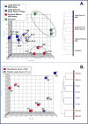
Results: Electrolyte and protein levels were relatively consistent among all serum-based PL lots, with only slight variations in glucose and calcium levels. All nine lots were as good as or better than fetal bovine serum in expanding MSCs. Serum-based PL stored at -80°C remained stable over 2 years. Quantitative cytokine arrays showed similarities as well as dissimilarities in the proteins present in serum-based PL. Greater differences in MSC gene expression profiles were attributable to the starting cell source rather than with the use of either PL or fetal bovine serum as a culture supplement.
Conclusion: Using a large-scale, standardized method, lot-to-lot variations were noted for industrial-scale preparations of serum-based PL products. However, all lots performed as well as or better than fetal bovine serum in supporting MSC growth. Together, these data indicate that off-the-shelf PL is a feasible substitute for fetal bovine serum in MSC cultures.
© 2017 AABB.
Conflict of interest statement
JAR, DFS, JP, AP, EB and PJin, have no conflicts of interest.
Figures


Similar articles
-
Characterization of platelet lysate cultured mesenchymal stromal cells and their potential use in tissue-engineered osteogenic devices for the treatment of bone defects.Tissue Eng Part C Methods. 2010 Apr;16(2):201-14. doi: 10.1089/ten.TEC.2008.0572. Tissue Eng Part C Methods. 2010. PMID: 19469694
-
Pathogen-free, plasma-poor platelet lysate and expansion of human mesenchymal stem cells.J Transl Med. 2014 Jan 27;12:28. doi: 10.1186/1479-5876-12-28. J Transl Med. 2014. PMID: 24467837 Free PMC article.
-
Platelet lysate as a substitute for animal serum for the ex-vivo expansion of mesenchymal stem/stromal cells: present and future.Stem Cell Res Ther. 2016 Jul 13;7(1):93. doi: 10.1186/s13287-016-0352-x. Stem Cell Res Ther. 2016. PMID: 27411942 Free PMC article. Review.
-
Effect of allogeneic platelet lysate on equine bone marrow derived mesenchymal stem cell characteristics, including immunogenic and immunomodulatory gene expression profile.Vet Immunol Immunopathol. 2019 Nov;217:109944. doi: 10.1016/j.vetimm.2019.109944. Epub 2019 Sep 21. Vet Immunol Immunopathol. 2019. PMID: 31563725
-
Human platelet lysate to substitute fetal bovine serum in hMSC expansion for translational applications: a systematic review.J Transl Med. 2020 Sep 15;18(1):351. doi: 10.1186/s12967-020-02489-4. J Transl Med. 2020. PMID: 32933520 Free PMC article.
Cited by
-
Acceleration of Translational Mesenchymal Stromal Cell Therapy Through Consistent Quality GMP Manufacturing.Front Cell Dev Biol. 2021 Apr 13;9:648472. doi: 10.3389/fcell.2021.648472. eCollection 2021. Front Cell Dev Biol. 2021. PMID: 33928083 Free PMC article. Review.
-
Chitosan hydrogel incorporated with dental pulp stem cell-derived exosomes alleviates periodontitis in mice via a macrophage-dependent mechanism.Bioact Mater. 2020 Jul 16;5(4):1113-1126. doi: 10.1016/j.bioactmat.2020.07.002. eCollection 2020 Dec. Bioact Mater. 2020. PMID: 32743122 Free PMC article.
-
Human Platelet Lysate Can Replace Fetal Calf Serum as a Protein Source to Promote Expansion and Osteogenic Differentiation of Human Bone-Marrow-Derived Mesenchymal Stromal Cells.Cells. 2020 Apr 9;9(4):918. doi: 10.3390/cells9040918. Cells. 2020. PMID: 32283663 Free PMC article.
-
Risks in Induction of Platelet Aggregation and Enhanced Blood Clot Formation in Platelet Lysate Therapy: A Pilot Study.J Clin Med. 2022 Jul 8;11(14):3972. doi: 10.3390/jcm11143972. J Clin Med. 2022. PMID: 35887736 Free PMC article.
-
Blood Plasma Derivatives for Tissue Engineering and Regenerative Medicine Therapies.Tissue Eng Part B Rev. 2018 Dec;24(6):454-462. doi: 10.1089/ten.TEB.2018.0008. Epub 2018 Oct 5. Tissue Eng Part B Rev. 2018. PMID: 29737237 Free PMC article. Review.
References
-
- Jochems CE, van der Valk JB, Stafleu FR, Baumans V. The use of fetal bovine serum: ethical or scientific problem? Altern Lab Anim. 2002 Mar-Apr;30(2):219–27. - PubMed
-
- Al-Saqi SH, Saliem M, Asikainen S, Quezada HC, Ekblad A, Hovatta O, et al. Defined serum-free media for in vitro expansion of adipose-derived mesenchymal stem cells. Cytotherapy. 2014 Jul;16(7):915–26. - PubMed
-
- van der Valk J, Brunner D, De Smet K, Fex Svenningsen A, Honegger P, Knudsen LE, et al. Optimization of chemically defined cell culture media--replacing fetal bovine serum in mammalian in vitro methods. Toxicol In Vitro. 2010 Jun;24(4):1053–63. - PubMed
-
- Kocaoemer A, Kern S, Kluter H, Bieback K. Human AB serum and thrombin-activated platelet-rich plasma are suitable alternatives to fetal calf serum for the expansion of mesenchymal stem cells from adipose tissue. Stem cells. 2007 May;25(5):1270–8. - PubMed
Publication types
MeSH terms
Substances
Grants and funding
LinkOut - more resources
Full Text Sources
Other Literature Sources

